Abdominal CT Scan with Contrast: What It Is and How It Works
February 29, 2024
Read More
Considered the leading cause of death in the US, heart disease affects nearly half the population. Compounded by the effects of COVID-19, public health authorities are bracing for a ripple effect, where heart disease numbers will escalate for the foreseeable future. Inevitably, there will be a rising need for heart imaging, of which there are many different types. Cardiac imaging is a crucial aspect of heart health to monitor the health of your heart and identify any potential issues.
In fact, a European study found that cardiovascular screening, including imaging and combined with treatment, may lower the risk of death, heart attack and stroke in men ages 65-69. The study found there was a greater benefit to screening if done before the age 70. In North America, heart imaging is a high priority for adults with comorbidities, especially those with diabetes, who are at an elevated risk of developing cardiovascular disease.
Here’s a look at the common types of cardiac imaging procedures and what you need to know about each of them.
An echocardiogram, or echo, is also known as a heart ultrasound or heart sonogram. Applying the same technology as an ultrasound, this non-invasive imaging test uses high-frequency sound waves to create an image of the heart. The test helps to visualize the size and shape of the heart, its chambers, and the movement of the heart’s walls. It also shows how blood flows through the heart valves, aiding in the detection of any structural problems. Echocardiograms are commonly used to diagnose heart disease, heart valve problems, and heart failure.
A cardiac computed tomography, also known as a CT, is a non-invasive imaging test that uses X-rays to create detailed images of the heart and blood vessels. A cardiac CT helps determine whether plaque or calcium deposits are present in the blood vessels and whether they are causing a blockage. It also provides information about the size and shape of the heart, the thickness of the heart walls, and the presence of plaque buildup in the coronary arteries. Cardiac CT scans diagnose general heart problems such as heart disease, heart blockages, and cardiac damage from a heart attack.
A single-photon emission computed tomography, also known as a SPECT, is a non-invasive nuclear imaging test that uses a special dye and a camera to create a detailed image of the heart. While most standard imaging tests show an organ’s structures, SPECT visualizes its function. It detects blood flow and blood distribution in the heart. It is particularly useful in diagnosing coronary artery disease, heart muscle damage, and heart blockages.
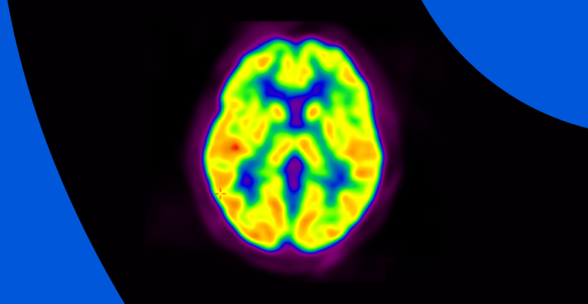
A cardiac positron emission tomography, also known as a PET, is a non-invasive heart imaging test that uses a special type of radioactive dye (a tracer) and a camera to create a detailed image of the heart. The test helps to detect blood flow, the distribution of blood in the heart, and the presence of heart disease. A heart PET scan can detect whether areas of your heart muscle are receiving enough blood, if there is heart damage or scar tissue in the heart, or if there is a buildup of abnormal substances in the heart muscle.
A coronary angiogram, also known as a left heart catheterization, is a minimally invasive procedure that uses a thin tube called a catheter to access the coronary arteries and visualize the inside of the heart. The test helps to diagnose heart disease, heart blockages, and other heart conditions. It is particularly useful in diagnosing problems with the heart’s blood vessels, such as blockages or stenosis.
A cardiac MRI, also known as a magnetic resonance imaging, is a non-invasive imaging test that uses a strong magnetic field and radio waves to create detailed images of the heart and blood vessels. The test provides information about the size and shape of the heart, the thickness of the heart walls, and the flow of blood through the heart and blood vessels. It is particularly useful in diagnosing heart conditions such as heart disease, heart muscle damage, and heart valve problems.
Finally, the last thing a person with heart problems needs is more stress. Waiting to receive imaging results can be a nerve-racking experience because you don’t know what you don’t know.
Fortunately, technology has made it easier for patients to access their heart imaging results and stay on top of their heart health. Most heart imaging test results are now available online through PocketHealth, giving patients fast and easy access to their records from anywhere and anytime.
With convenient access to imaging records, patients have the power to take better care of their heart health anywhere and anytime. Whether you are seeking an echocardiogram, a cardiac CT, or any other type of cardiac imaging, PocketHealth is a great resource for you to stay on top of your heart health and keep your heart in great shape.
Access My Records