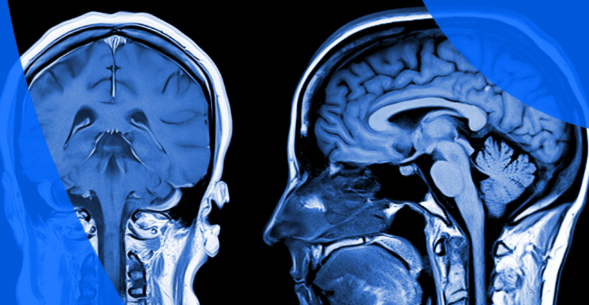
If you’ve had an MRI scan, you may be wondering what to expect when you get your results. As with many imaging reports, medical terminology and jargon can be confusing. This article will cover some of the language you might encounter in your results.
Your doctor will provide the medical interpretation of your MRI scan, but understanding terms like MRI views, signal intensity, contrast and enhancement may help you have a more enlightened follow-up appointment.
Magnetic resonance imaging (MRI) is a medical imaging technique that creates comprehensive images of your internal structures. It utilizes radio waves and magnets to view details of organs, blood vessels, muscles and bones. Here are examples of some of the medical conditions an MRI might help diagnose:
Possible abnormal findings in MRIs may show the following:
While it is understandable to feel nervous, abnormal findings don’t necessarily mean cancer or other serious conditions. It’s helpful to discuss any questions about your results with your doctor. Depending on the findings of your MRI scan, your healthcare provider may refer you for more imaging (such as a CT scan or ultrasound). Confirmation of your diagnosis may require a biopsy.
Report Reader is a valuable tool designed to empower you with a better understanding of your results. It translates complex medical terms into easy-to-understand language, enabling you to grasp the details and approach your next doctor’s appointment with confidence. The MyCare Navigator feature goes a step further by alerting you if your results indicate a need for follow-up, ensuring that you never miss an important discussion with your healthcare provider.
MRI with axial, coronal and sagittal views
MRI images are taken from three different angles across various planes of your body. This approach ensures the most accurate and detailed images. These cross-sectional images provide the radiologist with a detailed 360° overview and are referred to as the orientation. The 3 cross-sectional orientations are:
MRI scans showing the difference between T1 and T2 weighted scans
A T1-weighted MRI uses the rate of energy released as protons resume alignment (T1 relaxation) to create images. T1 images better show the anatomy of soft tissues of the body.
A T2-weighted MRI uses the oscillation of protons during the process (T2 relaxation) to create images. T2 images more accurately show the composition and placement of water molecules in tissue, highlighting cysts, fluids and swelling.
Together, T1 and T2 images provide doctors with complementary diagnostic information.
A low (hypointense) T1 signal compared to surrounding muscular or skeletal tissue often indicates bone lesions. The lesion area is generally compared to nearby structures and muscle to confirm the abnormality. A low T1 signal in bony areas can suggest increased water content and a decrease in normal fatty tissue. This can indicate conditions such as cancer, infections or trauma from an injury.
You may also come across terms like “diffusely low T1 marrow signal“. This typically indicates less fatty and more cellular or fluid-rich bone marrow. It could be due to anemia, infiltrative disorders like leukemia or metastatic cancer, inflammation, or changes from treatments like chemotherapy. The significance depends on the clinical context and should be assessed by a radiologist.
An increased or high (hyperintense) T2 signal can indicate a lesion in soft tissue. The tissue appears brighter than nearby areas, suggesting increased water content in these lesions. Possible causes of increased T2 signals include inflammation, cysts, abscesses, tumors and edema (areas of swelling).
Signal intensity is the level of brightness in an area of an MRI image. The variance in signal intensity can highlight the difference between an affected area and the surrounding tissue, leading to a successful diagnosis.
The level of signal intensity in an MRI image depends on the radiofrequency pulse used in the MRI machine, as well as the proton density and T1 or T2 qualities of various tissues.
What low signal intensity indicates depends on what pulse sequence is used. Fat, for example, is bright (meaning it has high signal intensity) in T1-weighted images. But fat and water and both are bright in T2-weighted images. Low signal intensity often means the area is darker compared to adjacent tissues, but there are many exceptions depending on the imaging method, such as T1 versus T2-weighted images.
For example, low signal intensity on T1-weighted scans can imply scar tissue, old hemorrhages or calcifications. On T2-weighted scans, this same low signal intensity can suggest certain tumors, dense tissue such as calcified lesions or iron deposits. It all depends on the imaging techniques and the makeup of the tissue or bone being scanned.
High signal intensity is often white on an MRI image. As with decreased intensities, indications vary based on imaging techniques and the areas of the body being scanned. For example, on T1-weighted scans, tissues with fat, areas of increased blood flow and inflammation or tumors can show up brighter on the MRI. T2-weighted scans might indicate cysts, other tumor types or areas of swelling and fluid retention.
Abnormal signals on MRI results can indicate a variety of issues. These signals can appear in both bony structures and soft tissue, showing as increased or decreased brightness in certain areas or deviating in appearance compared to baseline images, usually from past scans of the patient when available. For instance, an athlete may experience increased wear and tear as they age, which could result in abnormal MRI findings compared to previous scans.
The range of possibilities with abnormal signals is vast, including cysts, tumors, inflammation, infection, arthritis and multiple other conditions, many of which are benign. Because of this, try not to panic if you receive an abnormal signal in your report. Your doctor will discuss whether any further testing is needed and share their thoughts on potential diagnoses.
Fluid signals refer to fluid filled areas on the MRI. They typically look bright on T2-weighted scans but dark on T1-weighted scans. They are helpful for evaluating possible fluid-filled structures in the body, such as cysts, abscesses and edemas.
In MRI scans, a contrast agent is often used to enhance the visibility of blood vessels, organs, and soft tissues, making fine details more apparent. This agent, typically derived from gadolinium, is injected through an IV before the scan. By altering the magnetic properties of water in the body, the contrast agent improves the clarity and sharpness of the images, allowing for a more accurate diagnosis.
Enhancement refers to the increase of signal intensity due to the absorption of the contrast agent. Areas of increased blood flow, such as tumors, also have increased uptake of the contrast material.
Here is some terminology regarding enhancement that you may encounter in your final report and what it means:
Here’s a quick guide to understanding key MRI report terms: findings, indication, impression, and unremarkable. This section will help clarify what your MRI shows and what it means for your health.
The findings section of an MRI report details the observations made by the radiologist as they review the images. This part lists any abnormalities, changes, or notable features seen in the scanned area. It’s a descriptive section that records everything observed, even if it’s not necessarily related to the condition being investigated.
The indication section of your MRI report is a brief summary of the medical reason that necessitated the MRI in the first place. It is a way for the physician to communicate medical suspicions or symptoms to the radiologist who will interpret the images, providing them with a framework of your condition to keep in mind as they review your MRI.
In the context of an MRI report, the “impression” is the section where the radiologist provides a summary of the key findings and their interpretation. This part distills the complex imaging data into a clear, concise conclusion, highlighting any abnormalities or significant observations. The impression is typically the most important part of the report, as it gives the referring physician a clear understanding of what the MRI revealed and helps guide further diagnosis, treatment, or follow-up.
In MRI results, the term “unremarkable” means that the area being examined appears normal and shows no signs of abnormalities, disease, or any noteworthy findings. Essentially, it indicates that nothing unusual or concerning was detected in that specific part of the body.
Reading your brain MRI report is similar to what has already been discussed in this guide. However, it can be helpful to keep certain factors in mind as you review your results.
If you are having a repeat MRI to track progress on some condition or abnormality, your new MRI report will likely compare current findings to old findings.
Some possible findings you may see in an abnormal brain MRI include:
Remember, your MRI is a tool to help your doctor reach a diagnosis. It usually requires additional testing to pinpoint your condition, if any. It can be easy to worry about abnormal brain MRI results, but they are far more common than most people realize. Many irregularities turn out to be benign or easily treatable. Your doctor will be able to discuss any results that point to a normal versus abnormal brain MRI, as well as the next steps you may need to take.
Depending on the extent of your brain MRI, there will likely be a section describing these areas. If they are described as unremarkable, they are within normal expectations. The radiologist will indicate if there are abnormalities. Also, there are brain-adjacent terms you may come across, such as sella and parasellar. These are anatomical descriptions of the area around the pituitary gland, which is located at the base of the brain. Your doctor will be able to help you understand where these areas are if you find yourself uncertain.
You may come across terms like pachymeningeal enhancement, leptomeningeal enhancement, and intracranial enhancement when reviewing a brain MRI. Here’s what these terms mean:
These are some of the most important sections of your MRI report. They will include the radiologist’s determinations of your images. This may include specific measurements of any lesions or growths, descriptions of changes in your condition compared to previous MRI reports, or any other significant findings, theories or thoughts on diagnoses.
Patient viewing their MRI report on PocketHealth
Waiting for MRI results can be nerve-wracking. More than 50% of patients say that waiting for the results of an MRI or CT scan induces anxiety. Understanding the findings in your MRI report can help reduce that anxiety, as can gaining faster access to your finalized results with PocketHealth. As soon as your report is officially uploaded, you can safely access it anytime you need. While the doctor who ordered the MRI will explain your report at your next appointment, getting an early view can help you prepare and make the most of your time together.
With PocketHealth, you get fast and secure access to your MRI results from any device. You can easily view your MRI images and reports from any device, and even share them in diagnostic quality with specialists you want to obtain a second opinion. Jeanne found this especially important when she was abroad in Costa Rica and needed to see a local doctor for knee, hip and back pain. With all her old MRI scans and other imaging in one place, he was able to use them to give her a diagnosis and treat her pain, even when she was far from home.
PocketHealth Report Reader can help you better understand the findings in the radiologist’s report by providing easy-to-understand definitions of complex medical terms. MyCare Navigator detects and highlights follow-up recommendations and provides you with personalized questions to ask your doctor.
Updated: May 12, 2025
Trusted by more than 800+ hospitals and clinics.