Dr. Alexander Mansfield, Staff Radiologist at Headwaters Health Care Center in Orangeville, ON, explains what a bone density scan is and how it helps to detect bone disorders such as osteoporosis.
A bone density scan is a screening test to evaluate for decreased bone mass (known in medicine as osteopenia and osteoporosis) and help determine a patient’s fracture risk. A bone scan is also used to follow patients with low bone mass to assess the effectiveness of therapy which can include medications, exercise and other lifestyle changes.
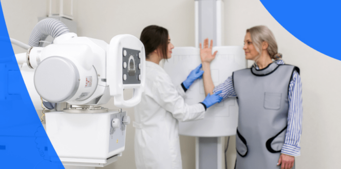
DEXA (dual energy x-ray absorptiometry) is the type of scan used to determine a
person’s bone density. Two x-ray energies are used. The density of the bone is measured by the strength of the x-ray beams passing through the body. It determines a patient’s bone mineral density in grams/square centimeter.
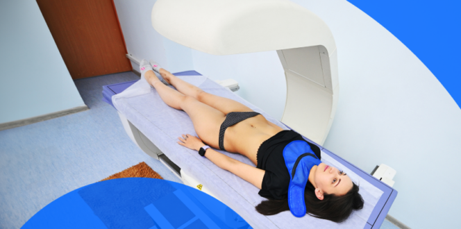
A bone scan determines the bone density of the spine and hip in grams/square centimeter.
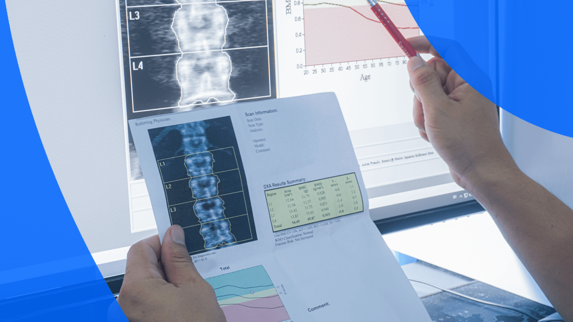
The bone density is then compared to a database of normals for a patient’s age and sex providing a T-score. This score gives a measure of how far above or below the normal average one is. The T-score can be expressed as a percentile or as a standard deviation from the normal average.
The T-score helps to determine a patient’s fracture risk along with other clinical information such as a history of a fragility fracture and steroid use.
Osteoporosis is a disorder characterized by decreased bone mass and an increased susceptibility to fractures. It is often related to increasing age and is a public health issue due to the number of related fractures. Women over 50 are at the highest risk with one in five developing osteoporosis.
Some other risk factors include a family history of bone fractures and osteoporosis, history of fractures past age 50, previous surgery to remove ovaries prior to menopause, lack of calcium or vitamin D, smoking, long-term use of certain medications, hormone imbalances or low body mass index or underweight.
A bone density scan looks like an x-ray. The radiologist will focus first on the spine to assess density. In a close-up, it looks like this:
A small amount of radiation is used to perform the scan – the lowest amount to achieve a diagnostic exam. Increased exposure to ionizing radiation can increase a patient’s risk of cancer, however, given the tiny amount used (equivalent to 3 hours of background radiation), the benefit of the exam outweighs the risk. More information about radiation safety can be found at www.imagewisely.org.
You can access your bone density scans along with other diagnostic imaging through PocketHealth. It should be available soon after the radiologist reviews the scans and writes a report, allowing you to easily share and send imaging to your healthcare providers in any medical practice or hospital network.
Access My RecordsPublished: March 7, 2023
Trusted by more than 900+ hospitals and clinics.