Suspect You Might Be Pregnant? What to Know About an Early Pregnancy Ultrasound

Early pregnancy can be a rollercoaster of physical symptoms and emotions. If you suspect you may be pregnant, it’s recommended to take a pregnancy test and schedule an appointment with an obstetrician. To confirm pregnancy, many providers perform blood tests and sometimes an early ultrasound to establish gestational dates. This guide will explain how ultrasounds are performed, how to prepare and what possible findings might mean.
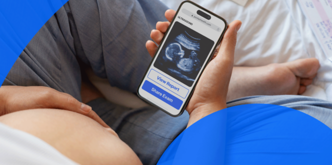
While this guide outlines potential findings, your doctor will provide your official diagnosis and next steps during your follow-up appointment. For those who prefer early access, PocketHealth offers secure, instant access as soon as your report is uploaded. This allows you to review your results and prepare questions for your doctor in advance.
Why get an early pregnancy ultrasound?
Getting an ultrasound is a normal part of pregnancy monitoring and the best way for your practitioner to keep tabs on your health and the development of the embryo. There are several reasons why you might have an early ultrasound:
- To establish the gestational age of the embryo: Measuring the size of the embryo using crown-rump-length helps narrow down the due date.
- To count the number of embryos: If there is more than one, an early ultrasound will attempt to discern if the embryos share a gestational and amniotic sac to determine the type of twins or multiples. Keep in mind, sometimes it is too early to see these details. The discovery of multiples may not come until a later ultrasound.
- To confirm the pregnancy is viable: A viable intrauterine pregnancy is safely implanted inside the uterus. An ectopic pregnancy (planted outside the uterus) or molar pregnancy (fertilized egg develops into a cyst-like mass) is, unfortunately, not viable.
- To detect the embryo’s heartbeat: The fetal heart rate will also be determined.
- To locate any sources of vaginal bleeding: If the patient shows potential complications, like bleeding or abdominal pain, an ultrasound can help determine the cause.
- To proactively monitor for potential complications: This is more applicable for those with underlying health conditions or a family history of such.
- To examine the cervix, ovaries and uterus: This examination can confirm if there’s an empty gestational sac or other signs of miscarriage or pregnancy loss. It also evaluates that these organs are reacting as expected during early pregnancy.
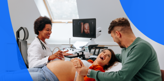
What to expect at an early pregnancy ultrasound?
Ultrasounds use high-frequency sound waves to create images of the patient’s organs and other internal structures. As these waves bounce off these areas, they help form images, which are then captured by a computer for measurement and evaluation.
During an ultrasound, the sonographer runs a transducer wand over conductive gel to send high-frequency sound waves into the body. The echoes created by the sound waves are processed into images. According to the U.S. Food & Drug Administration, ultrasounds are very safe, low-risk scans.
Types of pregnancy ultrasounds
There are two types of pregnancy ultrasounds:
- Transvaginal ultrasound: Transvaginal ultrasounds scan a smaller area more effectively to capture images by inserting a lubricated transducer wand into the vagina. Early in your pregnancy, you’re likely to receive a transvaginal ultrasound since the embryo is still very small at this point and an internal scan provides better visibility.
- Transabdominal ultrasound: Abdominal ultrasounds scan a wider area of your belly to capture images through the abdominal wall. The technician covers the area with a lubricating conductive gel to run the transducer through. This type of ultrasound is typically used when the fetus is more developed, starting around 10 or 11 weeks.
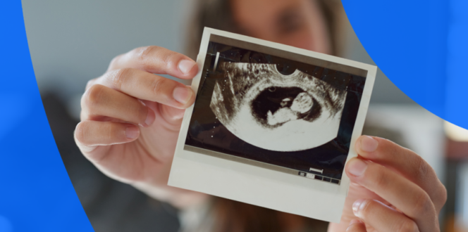
What can an early ultrasound pregnancy show?
What you can see in an early pregnancy ultrasound will depend on when you have it. Development is rapid in these early stages, so even half a week will make a difference in what the ultrasound can capture.
- 4 weeks: This early, it’s possible to see endometrial thickening, but it’s also possible to find no evidence of pregnancy yet.
- 5 weeks: The gestational sac, which surrounds the embryo in the early stages of pregnancy, becomes apparent.
- 5.5 to 6 weeks: The yolk sac is visible. Located inside the gestational sac, the yolk sac nourishes the embryo and is absorbed by the end of the first trimester.
- 5.5 to 6.5 weeks: At this stage, an ultrasound will distinguish the embryo, which is also sometimes called the fetal pole.
- 7 weeks: Though it might be possible earlier, at 7 weeks you’re likely to be able to make out the fetal heartbeat.
- 8 weeks: You can see the umbilical cord by 8 weeks, which passes nutrients and oxygen to the embryo.
- 9 weeks: Limbs are becoming distinct, so you should see defined arms and legs.
- 10 weeks: Developing hands and feet become more recognizable.
What the ultrasound will show depends entirely on the gestational age being accurate. Being off even by a few days can make a difference in whether or not the ultrasound can detect a heartbeat.
In early pregnancy you won’t see much detail. At 6 weeks, the embryo is only the size of a pea. From 8–11 weeks, you’ll be able to distinguish many more features, and in the 18-22 week anatomy scan, you’ll likely find out the sex of the baby.
How to prepare for an early ultrasound appointment
If this is your first prenatal ultrasound, it can help to know what to expect. Here are some tips for both before the scan and on the day of the procedure.
Before your scan
- Ask about any possible ultrasound fees: Depending on your insurance coverage, you may have some financial responsibility for the scans. It can help to call your doctor’s billing department before the appointment to make any necessary financial arrangements, such as payment plans, if applicable.
- Ask if you’ll need a full bladder: Many prenatal ultrasounds require one, but with early ones that use transvaginal methods, you may not. It helps to ask in advance so you know what preparations to make.
- Ask about morning sickness aids: If you have significant nausea (common in early pregnancy), consider asking your doctor about possible medications or recommendations. If you need a full bladder for your ultrasound, nausea may make drinking the required amount of water uncomfortable.
- Ask if you can bring someone with you: If you want someone present during the ultrasound, it helps to know the imaging clinic’s policies first. Many have restrictions on bringing small children or limiting the number of people allowed in the room.
The day of your scan
A pregnancy ultrasound isn’t a complex scan, but there are some things you can do to prepare, including:
- Bring a copy of your requisition with you: Your requisition specifies exactly which type of ultrasound exam you need. Having a copy with you will simplify the check-in process if your practitioner didn’t send it to the imaging clinic in advance. Also bring your health card and I.D.
- Drink plenty of water in the week before your appointment: Good hydration keeps your amniotic fluid clear, which means the ultrasound images are sharper.
- Book your appointment when your baby is most active: Movement will help the technician capture more images. Another suggestion is to have a cold and sugary drink just before your appointment.
- Arrive with a full bladder (if applicable): Sound waves and their echoes travel freely through liquid, so the sonographer can more easily capture images.
- Wear loose, comfortable clothing: Wear clothing you can easily take on and off.
An ultrasound technician is not legally allowed to discuss your scan results. But you can ask questions about the procedure itself, like:
- How long will this appointment last?
- Can I have someone with me during the ultrasound?
- Can I take my own photos or videos during my appointment?
- How can I access and share my images and results?
- When will my practitioner receive the results from you?
Getting your results
Patients experiencing their first pregnancy ultrasound are often excited about the results. Here is a quick overview of what to expect.
Who interprets my results?
Medical imaging is usually interpreted by a specialist called a radiologist, who reviews various scans to help evaluate and diagnose injuries and conditions. This information is then passed to your referring provider, who will incorporate their own assessment. For instance, if you have a history of certain conditions, they may use that data alongside the imaging for a more detailed analysis.
When will I get my results?
Pregnancy ultrasound turnaround times depend on the radiologist’s and your referring doctor’s schedules. It can sometimes take a week or more, especially if you receive them at your follow-up appointment.
With PocketHealth, you can get your results much faster. You’ll have secure access as soon as they’re uploaded, meaning you’ll likely see them before your upcoming appointment. This gives you time to review the findings and prepare for your next medical visit. MyCare Navigator is especially helpful for offering personalized insights and identifying any recommended follow-up steps within your report. It helps you formulate relevant questions to ask your doctor so you can make the most of your consultation.
Understanding my results
Although your doctor will explain your results to you, accessing your report before your appointment can give you an early preview. When reviewing your report, it’s common to find the medical terminology complex and confusing. PocketHealth Report Reader simplifies this by offering clear, straightforward definitions for medical terms—simply tap or click on any underlined words to reveal their meaning. Though your doctor will be the one to interpret your personal results, this guide will briefly discuss some common findings in pregnancy ultrasound reports.
Common findings in early ultrasound results
- Fetal biometry measurements: Fetal biometry involves measuring specific parts of the fetus, such as the crown-rump length (from the head to the bottom), femur length, etc. At this early stage, there will be fewer measurements, as the baby hasn’t developed enough yet, but the fetal pole and crown-rump length are visible.
- Gestational sac: Measurements of the sac help to determine possible due dates and gestational age.
- Heartbeat: The ultrasound may be able to pick up the fetal heartbeat, but it depends on how far along the pregnancy is.
- Location: This will confirm whether the embryo is implanted in the uterus, which is the only viable location.
- Number of embryos: There is no guarantee that this assessment will be accurate if the ultrasound is very early (around six weeks). Sometimes, one embryo may be hidden behind the other or just out of sight in the scan images. However, twins and multiples are usually detectable at this appointment.
Frequently asked questions
Here is an overview of commonly asked questions about early pregnancy ultrasounds.
How early can a pregnancy be seen on an ultrasound?
An ultrasound can determine a pregnancy as early as 4.5 weeks after your last period. Gestational age is calculated based on the first day of your last menstrual period and assumes a 28-day cycle.
How many more ultrasounds will I have in my pregnancy?
Ultrasounds are safe, trusted exams that give your practitioner the best information with which to monitor the progress of your pregnancy. Most practitioners will send you for an ultrasound at least twice:
- Weeks 6 to 8: A dating scan during your first trimester.
- Weeks 11-13: This is an optional third ultrasound that typically happens between weeks 11–13, to measure nuchal translucency and assess risk for any chromosomal abnormalities.
- Weeks 18 to 22: An anatomy scan to measure fetal growth and development.
If your pregnancy is deemed high-risk you might have more ultrasounds so your practitioner can continually monitor your progress. High-risk indicators include:
- Your age, if you are under 17 or over 35
- Experiencing ongoing health issues, such as diabetes, high blood pressure or a mental health condition
- Carrying twins or multiples
- Having had complications with a previous pregnancy
Which pregnancy ultrasound is most important?
All pregnancy ultrasounds are important, so it’s not entirely accurate to say that one is more important than the other. However, the two most standard ultrasounds are the viability/ dating scan, which determines the gestational age and due date, and the anatomy scan. This evaluates physical development, growth rates, organ development and other key baselines. Many patients only receive these two ultrasounds, though there are several reasons why patients would have additional ones.
Does everyone need an early pregnancy ultrasound?
Most doctors recommend an initial ultrasound within the first 6 to 12 weeks of pregnancy. It’s common for patients to have an “early ultrasound” between six to nine weeks to confirm gestational dates. However, many patients won’t have their first ultrasound until later in the first trimester, especially if they have known conception dates or if their doctor doesn’t think an earlier scan is necessary. It all depends on factors like patient preference, uncertain conception dates, complex medical history, etc.
When will an ultrasound show my baby’s sex?
Technically, an ultrasound as early as 11 weeks may reveal your baby’s sex. It’s not 100% accurate, but there’s a concept called the “nub theory” that plays a part. Babies have a “nub,” which is actually a genital tubercle. The theory suggests that if the nub is below 10 degrees from or parallel to the spine, the fetus is female. If the nub is angled more than 30 degrees from the spine, it’s thought to be male. Accuracy starts at around 70% at 11 weeks gestation and increases to nearly 100% by week 13.
That said, the ultrasound images and the fetus must be precisely positioned to make these determinations. At earlier scans, it can be challenging to obtain these precise angles as the baby is smaller and may be turned away or in the wrong position. Additionally, the results can occasionally be inaccurate. The most reliable ultrasound to determine the baby’s sex is the anatomy scan, which takes place between 18 and 22 weeks. By then, the baby is large enough for all anatomy to be clearly visible.
What if you can’t see the baby or hear a heartbeat?
Not hearing a fetal heartbeat at an early ultrasound can sometimes indicate a miscarriage. Additional testing will be performed to determine if this is the case. However, if the patient’s conception dates are inaccurate, the fetus may not be as far along and developed as originally thought. In this case, it may simply be too early to detect a heartbeat. This is a common occurrence, and in such cases, the doctor may recommend a follow-up viability ultrasound in seven to ten days to give the fetus time to grow and reassess.
Stay on top of your health with PocketHealth
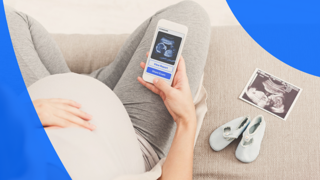
Pregnant patient checking results on PocketHealth
An early pregnancy ultrasound can be both exciting and nerve-wracking. Being prepared for your scan and follow-up appointment can help you feel more empowered from the very beginning. The more knowledge you have, the more empowered you’ll be at every step of your pregnancy journey, especially as you navigate those early days of pregnancy.
PocketHealth makes it simple to keep track of your medical reports and prenatal scans. All of your vital imaging is in one protected location and can be accessed anytime. When used in conjunction with your medical provider’s professional advice, it is a powerful tool for organizing and understanding your health’s progress. You can even use this platform to share your ultrasounds with family and friends throughout your pregnancy.
Published: August 3, 2023
Trusted by more than 800+ hospitals and clinics.