Baker Cysts: Causes, Symptoms, Diagnosis and Treatment

A Baker cyst, which develops behind the knee, is often the result of a knee problem rather than the cause of the problem itself. This article will examine what a Baker’s cyst is, how it is diagnosed, and how to understand the results.
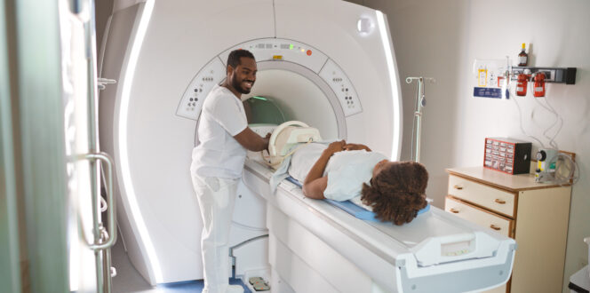
Baker cysts are often confirmed with internal imaging techniques such as MRIs, ultrasounds or X-rays. When you have medical images taken, you’ll receive your results at a follow-up appointment with your doctor. With PocketHealth, you can get your results more quickly, with secure, early access to your images as soon as they’re released by the radiologist.
What is a Baker cyst?
A Baker cyst is a thin-walled, fluid-filled sac that develops in the back of your knee. It is sometimes referred to as a popliteal cyst, or a popliteal synovial cyst. It might be apparent to the eye or touch, but many Baker cysts are asymptomatic and are only discovered when internal imaging is performed.
What causes a Baker cyst?
Knee joints are filled with synovial fluid to lubricate their movement and function. Injury or disease can occasionally allow excess fluid to ‘leak’ behind the knee and build up in a cyst. Baker cysts are not uncommon. They appear in children but are more frequent in adults.
Those most at risk of developing Baker cysts usually have another underlying condition such as osteo- or rheumatoid arthritis, or a tear to a ligament or the menisci, the pads which dispel the friction between the knee joint and the lower leg.
What are the symptoms of a Baker cyst?
Baker cysts can be entirely asymptomatic. You might not notice you have one until it is located by medical imaging. But if you do have symptoms, they may include:
- Lump: You might notice a lump behind your knee or feel a sense of fullness, especially when your leg is straightened.
- Pain: The area behind your knee might be uncomfortable or even painful to touch.
- Stiffness: Your knee might feel stiff or unwilling to fully bend, flex or straighten.
- Swelling: Your knee or leg might become swollen.
How is a Baker cyst diagnosed?
Baker cysts are frequently asymptomatic, though they can be discovered during a physical exam. If you do feel pain or swelling in the knee, however, you’ll likely be sent for medical imaging to confirm the diagnosis and ensure there are no other, more serious causes, such as an aneurism, blood clot or tumor.
Several different imaging techniques can be used to diagnose a Baker cyst. Which test you have will depend on your doctor, the availability of the imaging equipment and your circumstances and symptoms.
Ultrasound
Ultrasounds use high-frequency sound waves to produce live and still images of your internal soft tissues and structures. Ultrasounds can show if a lump behind your knee is full of fluid (cyst) or solid (mass).
X-ray
X-rays use low-dose radiation to capture images of the hard structures of your body. X-rays cannot distinguish cysts, but they can identify any damage to the bones in your knee joint that might cause a Baker cyst.
MRI
Magnetic resonance imaging scans (MRIs) use very strong magnets and radio waves to capture detailed images of your internal tissues and structures. They can help locate both the cyst and identify the underlying cause, such as a tear in the menisci.
How to prepare for your scan
When it comes to preparing for an ultrasound, X-rays or MRI, many of the general steps are the same, but some are test specific. For instance:
- Inform the clinic if you are pregnant: MRIs or X-rays may require some alteration in procedure if you are pregnant, so tell the hospital or imaging clinic when your appointment is booked.
- Check for special instructions: Read your requisition carefully and ask the clinic to clarify if there is anything you’re unsure of. Some MRIs require the use of a contrast agent, which may necessitate you avoid taking some daily medications before your test.
- Wear comfortable clothes: You’ll likely have to remove at least the bottom part of your clothes, so wear something you can easily take on and off.
- Bring your requisition with you: It contains important information for the imaging clinic staff. Also bring your health or ID card.
- Remove all jewelry and metal accessories: Metals can interfere with several types of imaging tests, so it’s best to leave all jewelry at home. This is particularly important with MRIs, as metals interfere with the magnetic imaging process. Inform your doctor if you regularly work with metals, have metal dental work that cannot be removed, or have any tattoos.
Getting your results
This section is an overview of your knee imaging results, including how long it takes to get them and how to understand them.
Who interprets my results?
A radiologist will interpret your scan results, and then forward the images and an accompanying report to your referring physician. Your doctor will share the results with you at a follow-up appointment.
How long does it take to get my Baker cyst scan results?
It can take up to a week or even longer to get your results, depending on the radiologist and when you can get an appointment with your own doctor.
PocketHealth gives you faster access to your medical images and reports. Once you have your results, MyCare Navigator can help you prepare for your appointment by providing you with personalized questions to ask your doctor and highlighting any recommendations in your report.
Understanding my results
Medical imaging reports contain complex medical terminology. Report Reader gives you easy-to-understand definitions of medical terms, so you can be fully informed going into your follow-up. Your doctor will be the one to officially explain your imaging report, but this next section explores general scan findings and what they may look like.
What a Baker cyst looks like on an ultrasound
Ultrasound images appear in shades of black, gray and white. The denser the tissue, the brighter it will appear. A cyst looks like a black, small fluid filled sac.
What a Baker cyst looks like on an X-ray
An X-ray won’t show the cyst itself, but it can expose conditions in the knee that may have led to the cyst, such as signs of osteoarthritis or damage from an injury.
What a Baker cyst looks like on an MRI
On an MRI, a cyst typically looks like a rounded fluid filled shape, usually lighter in color than the surrounding area.
How is a Baker cyst treated?
If you have no symptoms, you may not require treatment. Many Baker cysts resolve themselves without any intervention. However, if the cyst is very large or painful, you might receive one or more of the following treatments:
- Medications: Regular non-steroidal, anti-inflammatory drugs (NSAIDs) like acetaminophen (e.g. Tylenol) or ibuprofen (e.g. Advil) reduce swelling and discomfort. A steroid injection might be used to alleviate pain and swelling in more severe cases.
- RICE method: Rest, icing, compression, elevation: this method can also reduce inflammation and relieve pain.
- Arthrocentesis: A doctor might drain the synovial fluid with a needle in this process, which is also called joint or needle aspiration.
- Surgery: Surgery is rarely needed to remove a Baker cyst. However, it might be used as a last resort if other treatments are ineffective.
Even more importantly, the underlying condition that caused the Baker cyst must be treated. That treatment will depend on the nature of the disease or injury in question. For instance, if you have rheumatoid arthritis, you might benefit from certain medications and/or physical therapy. If an injury to the menisci caused the cyst, you may require arthroscopic surgery to fix the tear. Treating the underlying cause will keep the Baker cyst from returning.
Frequently asked questions
Here are some of the common questions people have about Baker cysts.
What should you avoid when you have a Baker cyst?
Baker cysts can sometimes be aggravated by high-impact aerobic activities, such as running, so it’s best to avoid those activities until you have a treatment plan.
Do Baker cysts need to be removed?
Generally, Baker cysts do not need to be removed. In fact, many of them dissolve on their own, with no intervention.
What happens if a Baker cyst stays untreated?
Many Baker cysts will resolve themselves without treatment, especially if the underlying condition that caused them is addressed.
In rare cases, a large, untreated Baker cyst can swell to the point that it blocks blood flow, compresses nerves, or ruptures. A ruptured cyst can mimic deep vein thrombosis, a potential serious disorder. Early diagnosis and treatment will help prevent those negative health outcomes.
Can a Baker cyst turn cancerous?
No, Baker cysts are benign. They’re filled with synovial fluid, the lubricating fluid that keeps the knee joint working smoothly, and they are often the result of another underlying condition such as a knee injury or arthritis.
Take control of your health with PocketHealth
PocketHealth makes it easy to keep track of all your medical images and reports. If you need one or more imaging tests to diagnose a Baker cyst, you can enjoy peace of mind knowing all your results can be accessed on any device.
See, share and store your medical images in full confidence with PocketHealth.
Published: January 9, 2025


Trusted by more than 800+ hospitals and clinics.