20 Weeks Pregnant: What to Expect from Your Anatomy Ultrasound
Generally performed between 18-22 weeks, the anatomy ultrasound checks for several congenital abnormalities and assesses the physical development of the fetus. This article will explore why you’ll have an anatomy ultrasound around 20 weeks, what the anatomy scan can detect, how to prepare for your ultrasound and how to access your images and report.
Why you may need an ultrasound at 20 weeks
You will usually have at least two prenatal ultrasounds, one in early pregnancy during the first trimester and the other during the second trimester. The second ultrasound happens between weeks 18-22. It is often called the anatomy scan, since doctors use it to monitor the development of internal organs and check that growth is progressing normally.
The anatomy scan is also occasionally referred to as an anomaly scan, as it can detect several health conditions, including:
- Abdominal defects, such as gastroschisis (intestinal extrusion) or omphalocele (extrusion of other abdominal organs)
- Anencephaly (underdevelopment of brain and skull)
- Cleft lip
- Congenital heart defects
- Hernia of the diaphragm or other muscles
- Kidney issues such as renal agenesis (missing kidneys)
- Skeletal dysplasia
- Spina bifida
- Trisomy 21 (indicator for Down syndrome)
- Trisomy 18 (indicator for Edwards syndrome)
- Trisomy 13 (indicator for Patau syndrome)
What can an anatomy scan detect?
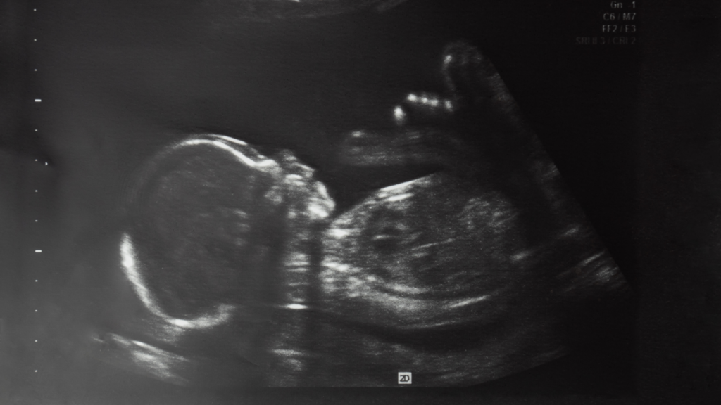
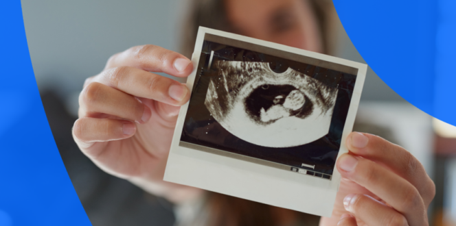
An ultrasound showing a 20-week fetus
The 20-week anatomy scan takes many important measurements to assess physical development as well as any congenital abnormalities. It is also generally the first point at which it is possible to identify the sex of the fetus, if you choose to.
- Brain: The sonographer will examine the size and shape of the brain and will assess brain fluid, looking for cysts or other abnormalities. Finding a cyst does not necessarily indicate a problem: in most cases, cysts resolve themselves by week 28.
- Heart: The anatomy scan will monitor how the heart looks and functions, assessing its size, positioning and the development of all four chambers. The technician will also be able to offer reassurance that fetal heart rate falls in the normal range (120-180 bpm).
- Limbs: The ultrasound technician will check that the bones in arms and legs are developing properly, along with the development of hands and fingers, feet and toes.
- Organs: Additionally, the scan will monitor the development of other organs like the bladder, kidneys, lungs and stomach. The sonographer will also gauge the health of the abdominal wall and diaphragm.
- Sex: At this point in the second trimester (18-22 weeks), the external genitalia are usually developed enough to identify, though not always. And depending on the position of the fetus it is possible that the genitals may not be visible.
- Spine: The sonographer will inspect fetal spine development, to ensure the vertebrae are well aligned and that the epidermal layer completely covers the spinal cord.
- Other: If the fetus’ position allows, the anatomy scan will also check the face for evidence of cleft lip. The ultrasound technician also uses this ultrasound to assess maternal health, checking the cervix, ovaries and uterus. They will also measure the amniotic fluid, check that blood flow is unhindered in the umbilical cord and ensure the placenta does not cover the cervix (placenta previa).
How to prepare for your 20 week ultrasound
An anatomy scan does not require too much preparation. Check with the imaging clinic for any special instructions when you book your appointment and be sure to bring your health or insurance card.
- Bring the requisition from your doctor: It contains important information for the imaging clinic.
- Arrive with a full bladder if required: Sound waves travel well through liquid: a full bladder helps the sonographer capture the clearest images. But at 20 weeks, it may not be required. Check with your clinic before your scan.
- Wear comfortable clothes you can easily adjust or remove. You may not need to remove your clothing, but you’ll need to expose your entire midsection.
Get fast access to your ultrasound results
What to expect at a 20-week ultrasound
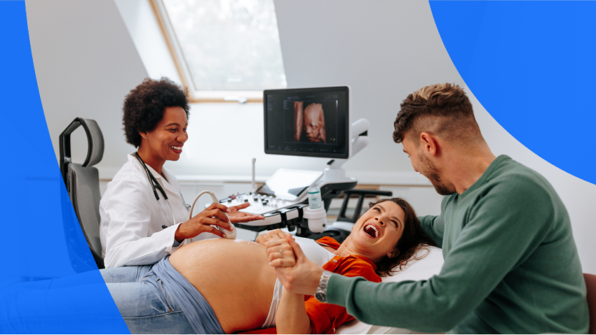
A 20-week anatomy scan is a safe, non-invasive procedure which does not involve the use of radiation. After you lie down on the exam table with your abdomen exposed, the technician will coat your abdomen in sound-conducting gel, then run a sound wave emitting ultrasound wand over the surface of your skin.
To capture the best images, the sonographer might move the transducer at different angles or pressures. You may need to change positions or to walk around during the process, or even be asked to drink something sweet to make the fetus more active.
How long will my ultrasound take?
The 20 week anatomy scan usually takes at least 45 minutes.
When is my next ultrasound after 20 weeks?
If the results of your 20-week anatomy scan are normal, you might not need to have further tests or imaging during your pregnancy. If your pregnancy is considered high risk, or there are any findings that need further exploration, you might require further ultrasound imaging.
How accurate is the baby’s gender at 20 weeks?
By 20 weeks, external genitalia are typically (though not always) well-enough developed for the sonographer to distinguish a labia or penis, but only if the fetus is facing the right way. Let the technician know ahead of time whether or not you wish to be informed of the fetus’ sex.
How big is the baby at 20 weeks?
At your 20 week ultrasound, the fetus will be 25-28 cm long from head to foot, with a crown-rump length of 16-17 cm.
What can you see on a 20-week pregnancy ultrasound?
The limbs and digits should be distinct, as the head. Facial features will be recognizable. You’ll likely be able to see hair and, if the fetus’ position permits, genitals.
How quickly will you get your 20 week pregnancy report and ultrasound pictures?
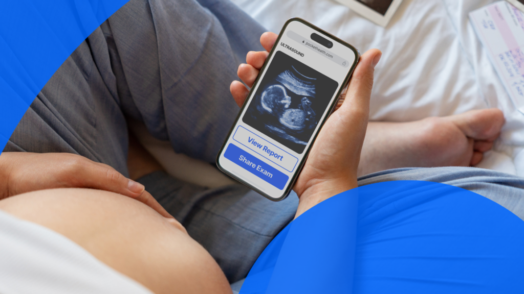
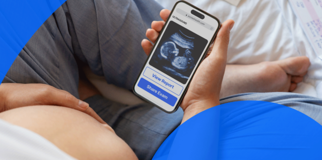
Pregnant person using PocketHealth to view their ultrasound
The clinic will forward your 20 week anatomy scan images and report to your referring physician, who will discuss them with you at a follow-appointment. That process can take just a few days or well over a week. That can be frustrating if you’re eager to see proof of your pregnancy’s progress.
With PocketHealth, you can get early access to your 20 week anatomy scan images and report, often as soon as they’re released by the imaging clinic. This means you can instantly share the joy and excitement of your baby’s development with friends and family, showcasing detailed ultrasound images that capture every precious moment.
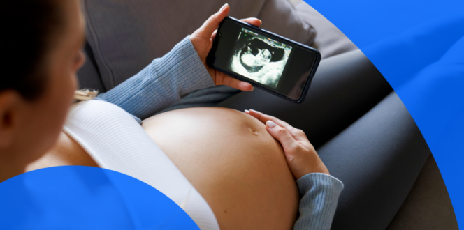
Feel confident throughout your pregnancy journey
With PocketHealth, you can easily access your pregnancy ultrasounds and reports on any device, wherever you are. At 20 weeks, you’re halfway through your pregnancy, and PocketHealth helps you track your baby’s growth and progress, allowing you to plan for the exciting journey ahead.
Report Reader simplifies complex medical terminology, making it easier to understand your report, while My Care Navigator provides personalized questions and highlights important follow-up recommendations, empowering you to feel informed and confident for your next appointment.
Published: October 4, 2024
Trusted by more than 800+ hospitals and clinics.