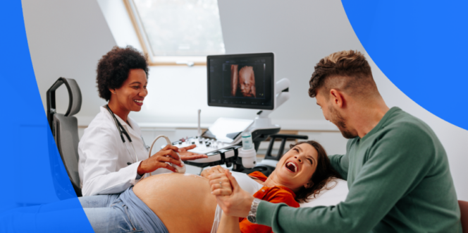
When you’re expecting, ultrasounds are a big part of your pregnancy journey. Not only do pregnancy ultrasounds help you and your healthcare team learn about your baby’s health and development, but they’re an excellent way to share your experience with family and friends.
It’s normal to experience a range of emotions around ultrasound appointments. Whether you’re nervously awaiting results or excitedly anticipating your next scan, the more you know about prenatal ultrasounds and why they’re performed, the more prepared you’ll feel at your appointments. In this article, you’ll learn the ins and outs of your upcoming ultrasounds, including:
First things first! Ultrasounds use sound waves to capture live images of your baby—pictures you can see right away on the ultrasound monitor. A pregnancy ultrasound is sometimes called a sonogram.
Your practitioner will send you for an ultrasound at least twice during your pregnancy. They’ll want to confirm the pregnancy by observing a heartbeat, determine gestational age and sex of your baby, as well as monitor growth and development. If you have a complicated or high-risk pregnancy, you might be sent for ultrasounds more frequently. This lets your practitioner check for development issues or screen for complications. A pregnancy may be classified as high risk if there is any reason to believe the baby may not be carried to full term or if health concerns are likely to arise.
A pregnancy might be considered high risk if a woman:
Yes, when performed by a trained sonographer, ultrasound technician, or radiologist according to Health Canada and the U.S. Food & Drug Administration. 2D, 3D, 4D and Doppler ultrasounds use the same sound-wave technology to safely capture images inside your body. Unlike X-rays, ultrasounds don’t use radiation to capture images. Only practitioners trained to perform fetal ultrasounds can give you a pregnancy ultrasound.
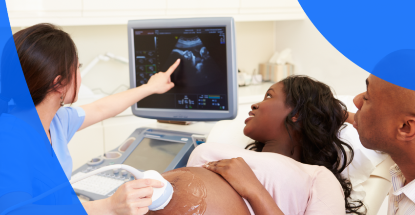
During an ultrasound, a physician, ultrasound technician, or trained sonographer uses a hand-held device called a transducer. The transducer creates sound waves (too quiet for human ears to hear) that travel painlessly through your body to your baby. An ultrasound machine then detects the sound waves and uses them to create an image of the exact position and shape of your baby. Ultrasounds typically produce a flat, 2D image of your baby, but 3D ultrasounds can give you a more detailed view.
3D and 4D ultrasounds can be very exciting for expectant parents because they can show facial expressions and allow you to see what your baby looks like. These types of ultrasounds can also be used in conjunction with 2D diagnostic ultrasounds to showcase certain facial abnormalities, like a cleft palate.
There are five types of pregnancy ultrasounds:
A pregnancy ultrasound is the best way for your practitioner to monitor your health and the development of your baby. Some common reasons your practitioner may send you for an ultrasound include:
Practitioners also use ultrasounds to help screen for specific health conditions or in response to certain events, like vaginal bleeding. Your practitioner may send you for an ultrasound to check for:
Your practitioner may send you for an ultrasound at any time during your pregnancy. Pregnancy ultrasounds typically happen only during the first and second trimesters unless the pregnancy is considered high-risk or complications develop.
Your healthcare practitioner will likely send you for your first pregnancy ultrasound no earlier than 6 weeks since a heartbeat can’t be easily detected before then.
An early pregnancy ultrasound can show your doctor how the embryo is developing and how your body is responding. By 5 weeks it’s possible to view the gestational and yolk sacs, and by 7 weeks, you’ll be able to see the embryo inside the amniotic sac.
The yolk sac nourishes your baby and can be an important indicator of fetal health early in pregnancy. However, it can only be seen in early pregnancy ultrasounds, since it gets absorbed into your body after the development of your placenta by the end of the first trimester.
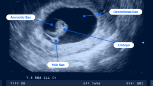
Ultrasound image at 7 weeks pregnant
If your pregnancy is low risk, your practitioner will likely send you for two ultrasounds: One during the first trimester and one during the second trimester. If your pregnancy is high-risk or complicated, your practitioner may send you for further ultrasounds. Here’s a general guide about what to expect.
Your practitioner may also recommend further ultrasounds if you carry your baby longer than 40 weeks or as part of diagnostic tests for specific pregnancy concerns, such as CVS and amniocentesis.
Your practitioner may also suggest you go for an elective 3D ultrasound to check on any physical abnormalities already detected in a routine 2D ultrasound.
Pregnancy ultrasounds require some preparation, and you may feel more comfortable attending your appointment if you know in advance what type of ultrasound you’ll be having. Transvaginal ultrasounds are typically done early in pregnancy because the embryo is so small, and this ultrasound can capture a better image from the vaginal canal.
You can prepare for your pregnancy ultrasound by:
If you’re worried about your ultrasound appointment, asking your practitioner or sonographer questions in advance can help you feel more prepared. Write down your questions ahead of time, including:
Every ultrasound you attend gives your practitioner important information about your pregnancy, so remember to ask questions at your follow-up appointment, too.
You can discuss your ultrasounds with your practitioner, but your sonographer cannot answer questions during your ultrasound. You may want to ask your practitioner these important questions at your follow-up appointment:
We’ve heard that many patients want to see their ultrasound images and reports as quickly as possible. With PocketHealth, you can quickly and easily access and share your pregnancy ultrasound images and review your reports—often before seeing your practitioner for a follow-up. Access your records here.
The PocketHealth platform enables you to securely access, share and store your imaging and other health information all in one place. It also enables you to share images with family and friends, so they can see firsthand how your baby develops and changes throughout your pregnancy journey.
Because pregnancy ultrasound terminology can be confusing, PocketHealth’s Report Reader is there to help. Report Reader makes it easier to understand terms in your ultrasound report and feel more prepared when speaking to your pregnancy care practitioner.
Pregnancy ultrasounds are a valuable medical tool that gives your practitioner important information about the growth of your baby, so understanding your pregnancy ultrasounds can reduce any stress around the unknowns that come with preparing for these appointments.
It’s also important to be informed when reviewing your results and speaking to your practitioner. Knowing which questions to ask helps you become a more active participant in your pregnancy care, no matter what trimester you’re in. Understanding why your practitioner has requested a pregnancy ultrasound can help you happily anticipate your next appointment and give you peace of mind throughout every step of your pregnancy.
Learn more about how to use PocketHealth to access and share your pregnancy ultrasound records.
Published: June 29, 2023
Trusted by more than 900+ hospitals and clinics.