Polycystic kidney disease ultrasound: How to prepare and what to expect

Polycystic kidney disease (PKD) is a genetic disorder that causes cysts to form in the kidneys. These cysts can damage the kidneys and may eventually lead to kidney failure, which could require dialysis or a kidney transplant. Approximately 600,000 people in the U.S. are affected by this condition, with equal risk across all sexes and genders. PKD is also responsible for about 5% of kidney failure cases.
This article provides an overview of PKD, focusing on the role of diagnostic ultrasounds in assessing and diagnosing the disease. You will learn how this imaging method is used and what to expect during your appointment.
What is polycystic kidney disease?
PKD is a genetic disorder, though it can be caused by various genetic mutations rather than a single one. Fluid-filled sacs, known as cysts, form inside the kidneys, also sometimes called renal cysts. In many medical conditions, cysts are harmless and may either be easy to treat or resolve on their own.
In the case of polycystic kidney disease, however, these cysts can damage the kidneys and cause them to enlarge significantly. A healthy kidney typically weighs about a third of a pound and is roughly the size of a fist. In contrast, kidneys affected by PKD can sometimes grow as large as a football and weigh up to 30 pounds each. There are two main types of PKD, which will be discussed next.
What is autosomal dominant PKD?
Autosomal dominant PKD (ADPKD) is the most common type of the disease, accounting for about 90% of all cases. Dominant Polycystic kidney disease occurs when one of the patient’s parents passes on the genetic condition, giving the patient a 50% chance of inheriting this form of PKD. Symptoms usually start in middle adulthood, but they can also start as soon as childhood.
What is autosomal recessive PKD?
The other form of PKD is autosomal recessive polycystic kidney disease (ARPKD), and it is considerably rarer than ADPKD. This type occurs when both parents pass on the gene, giving the patient a 25% chance of inheriting the condition. Symptoms may not appear until later in childhood or adolescence, but they often begin shortly after birth. In some cases, the condition is diagnosed while the baby is still in the womb. While ARPKD tends to be more severe, significant medical advancements have greatly improved lifespans, quality of life and overall outlook for affected individuals.
Potential complications of PKD
There are various potential complications of PKD, though these can vary by individual and the severity of their condition. Some complications that some patients may experience include:
- Cysts in other body parts: Besides the kidneys, PKD can cause liver cysts and cysts in the ovaries, bowel, spleen, pancreas, heart or brain. While most of these growths are not serious, cysts in the heart may affect the valves, leading to heart murmurs. Cysts in the brain can sometimes result in an aneurysm, which is something many PKD are regularly screened for, in case intervention is needed.
- High blood pressure: For many patients, high blood pressure may be the first sign of PKD.
- Increased risk for kidney stones and UTIs: PKD can raise the risk of urinary tract infections and kidney stones.
- Gastrointestinal issues: Examples include feeling prematurely full when eating, bloating and abdominal pain.
What causes polycystic kidney disease?
Most cases of PKD are caused by a genetic mutation in the PKD1, PKD2 or PKHD1 genes, which are passed down from the patient’s parent or parents. Rarely, this gene mutation occurs spontaneously, meaning it mutates randomly in the patient and was not inherited or passed down. Only about 10% of PKD patients develop this mutation spontaneously.
Regardless of how the mutations occurred, there is a 50% chance that someone with this condition will pass it to each of their offspring. Additionally, if a patient has autosomal dominant PKD (ADPKD), they are highly likely to manifest some symptoms or signs of the condition at some point in their lifetime. The severity of their condition, however, may vary. Some patients experience more severe cases of the disease, while others may only have mild symptoms. For patients with a family history of this mutation, if they are tested and found not to have it, they will not be at risk of passing it along to their children.
What are the symptoms of polycystic kidney disease?
Many patients go years without any symptoms of PKD. That said, here are some common symptoms of the condition:
- Feeling full in the abdominal area
- A history of kidney stones
- Frequent kidney or urinary tract infections
- Enlarged abdomen due to the increased size of the kidneys
- High blood pressure (hypertension)
- Pain in the back, side, or abdomen
- Frequent headaches
- Bloody urine
- Kidney failure
It’s important to note that experiencing some of these symptoms does not necessarily mean you have PKD. There are many other possible causes for these symptoms. It’s important to discuss any concerns with your doctor, who can help determine the cause and recommend next steps.
How is polycystic kidney disease diagnosed?
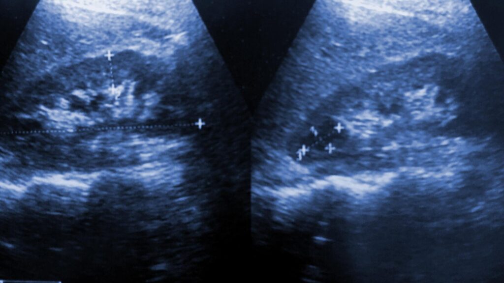
Ultrasound of a kidney cyst
There are generally multiple components to a PKD diagnosis. Here is a brief overview of how a diagnosis is typically reached.
Genetic testing
Despite PKD being caused by genetic factors in the majority of cases, genetic testing via blood sample is not always the first course of action for diagnosis. The test can be expensive for some patients and in 15% of cases, it doesn’t detect the condition even if the patient has it. However, genetic testing can be helpful in the following circumstances:
- Family history of the disease: Due to the increased risk, the patient may want to see if they have the genetic mutation as well.
- Imaging tests are inconclusive: If medical scans don’t clearly indicate PKD, genetic testing can be another way to help narrow down the diagnosis.
- Family planning purposes: If a patient is planning to start a family, genetic testing may be considered, especially if there is a family history of the disease.
Polycystic kidney disease ultrasound
Ultrasounds are generally the first method used to diagnose PKD. This imaging technique uses high-frequency sound waves produced by a hand-held device called a transducer. The ultrasound technician moves the transducer externally across the kidney area, and the sound waves bounce off internal structures and organs, echoing back to create images that can be captured and analyzed. The size of the kidneys and the presence of a polycystic kidney or kidneys can usually be detected, though this depends on the progression and severity of the disease.
Other imaging
Other imaging methods can also help with diagnosis, though they tend to be more expensive and may be harder to schedule. MRIs (magnetic resonance imaging) use radio and magnetic waves to create highly detailed images of internal anatomy. CT scans (computed tomography) use X-ray technology to produce detailed cross-sections of internal structures. Both can detect very small cysts that ultrasounds may miss, but generally, ultrasounds are a highly reliable, non-invasive diagnostic method that is easier to schedule.
How is a PKD ultrasound performed?
Because most PKD patients have an ultrasound for the first diagnostic test, it can be helpful to know what to expect. Here are the steps for most kidney ultrasounds:
- You will most likely be asked to arrive at your appointment with a full bladder. This is because a full bladder improves the transmission of sound waves and helps tilt certain organs into a better position for imaging.
- You will be asked to lie on an exam table, and a conductive gel will be applied to your skin over the area of your kidneys. The technician will slowly move the transducer over this area, capturing multiple images to get specific views and angles of your kidneys.
- You may be asked to hold your breath for short periods, as this can help improve imaging angles. Additionally, you may be asked to move to your side for some of the imaging.
- The ultrasound technician may also ask you to empty your bladder halfway through the scan and return for additional imaging. This allows them to compare the kidneys and bladder when full and empty.
- After the scan, you may return to your usual routine and go about your day.
How to prepare for a PKD ultrasound
Here are some helpful tips to prepare for your ultrasound appointment:
- Bring your requisition or ultrasound orders: The imaging clinic may already have these, but if they don’t, it could save you time at check-in.
- Bring your health card or insurance card: Bring your identification, as this is commonly required.
- Review any preparatory instructions in advance: Examples include full bladder preparation or directions to the facility.
- Wear a two-piece outfit: While some facilities may require a hospital gown for the scan, most will simply move the patient’s shirt slightly to access the kidney area. Wearing something comfortable is also helpful, and avoid wearing metallic items if possible.
Getting your results
Turnaround times for imaging results can vary widely depending on the facility and your doctor’s availability. Often, patients wait a week or more and receive their results during a follow-up appointment. With PocketHealth, you don’t have to wait as long—your results are securely accessible as soon as the report is available, allowing you to review them often before your follow-up visit.
Your imaging results are interpreted by a radiologist—an expert in medical imaging—who carefully reviews your ultrasound and provides a detailed report of any findings. This report is then sent to your referring doctor, who, with a deeper understanding of your medical history, can offer further insights and recommendations.
To better understand your imaging reports, Report Reader provides clear, straightforward definitions for medical terms. Simply tap or click on any underlined words to reveal their meaning. This is paired with illustrations and highlights of anatomy in your imaging to help you better understand your results and prepare for follow-up appointments.
Frequently asked questions
Here are some commonly asked questions regarding polycystic kidney disease and ultrasound scans.
How is acquired cystic kidney disease different from PKD?
Acquired cystic kidney disease (ACD) has similar symptoms to PKD, such as multiple cysts, but they have different causes. PKD is caused by genetic factors, while ACD is usually a result of chronic kidney disease, a condition with multiple potential causes. Additionally, ACD patients typically have normal or smaller-sized kidneys, whereas PKD patients often have enlarged kidneys. Finally, cysts generally do not form in other areas of the body in ACD patients.
Are there any risks from a kidney ultrasound?
A kidney ultrasound is a non-invasive and safe imaging procedure that can be performed repeatedly with no adverse effects. The only potential issue for some patients is slight discomfort from having a full bladder, particularly in more advanced stages of the disease. In such cases, speak with your physician or ultrasound technician, as adjustments may be possible.
At what age do patients usually start showing symptoms of PKD?
The age at which a patient with PKD begins to show symptoms can vary. For autosomal dominant PKD, symptoms are more commonly seen between the ages of 30 and 40, though there are exceptions where they appear earlier. For autosomal recessive PKD, symptoms are usually present before or shortly after birth.
Will a kidney transplant cure PKD?
A kidney transplant doesn’t cure the underlying cause of PKD, as it is usually genetic. This means other complications, such as the risk of brain aneurysms, would still need to be medically managed. There is also the potential for the new kidney to eventually become symptomatic. That said, many patients find their quality of life greatly improves after a kidney transplant, and many can live for years without complications with the new kidney, if they experience any at all.
Getting support
A PKD diagnosis can be overwhelming, and finding support is important. Several resources are available for patients with PKD. One of the first places to start is your doctor’s office, as they will likely have information on local support resources. On a broader scale, here are some online support options that may also help:
- PKD Foundation
- American Association of Kidney Patients
- National Kidney Foundation
- PKD Foundation of Canada
Take control of your health with PocketHealth
PocketHealth makes it simple to keep track of your kidney scans. All of your vital imaging is in one secure location and can be accessed online anytime. Reports can also be easily shared with other physicians in your care team, if needed. Additionally, it makes it easy to track health changes over time, such as with repeat imaging like ultrasounds. When used in conjunction with your medical provider’s professional advice, it is a powerful tool for organizing and understanding your imaging results and your health.
PocketHealth MyCare Navigator gives personalized insights into your health and identifies any recommended follow-up steps. This feature can also generate individualized questions to ask your doctor based on the findings in your report, ensuring you make the most of your consultation. A PKD diagnosis may feel overwhelming, but working closely with your providers can give you the best chance of managing your condition and protecting your kidney health.
Published: April 8, 2025


Trusted by more than 800+ hospitals and clinics.