Second only to skin cancer, prostate cancer is the most common cancer type for patients assigned male at birth (AMAB). It tends to develop more in older patients, with one in ten receiving this diagnosis at some point in their lifetime. African American AMAB patients also tend to have a higher rate of developing this disease.
This article will discuss diagnostic methods for prostate cancer such as medical imaging and other forms of testing. It will also give a brief overview of the disease and highlight educational and support resources that some may find helpful.
The prostate is an important part of the male reproductive system. This small gland produces fluid that mixes with sperm to form semen. Prostate cancer occurs when cells within the prostate grow and spread out of control, sometimes forming tumors or growths. When this happens, it can affect the health and function of the prostate and even lead to urinary issues. This is because the gland is located near the bladder and the urethra passes through the prostate.
While prostate cancer can be serious and is the second most common cause of cancer deaths in AMAB patients (behind lung cancer), mortality rates have significantly declined due to earlier detection and advancements in treatment. Most people who develop the disease are able to be cured as long as the condition is caught before it spreads. When it does spread (called metastasizing), there are still often treatments available to prolong the patient’s life.
Early prostate cancer cases may not cause obvious symptoms, but they are often detected through routine health screenings. When prostate cancer does become symptomatic, symptoms may include:
It is important to remember that there are numerous possible causes for these symptoms other than cancer. As AMAB patients age, their prostate can naturally enlarge, causing many similar symptoms. This is quite common and, while it may require treatment or adjustments, it is not usually life-threatening. Urinary tract infections are another possible cause of some of these symptoms, along with other benign conditions. It is recommended to consult with your doctor about any symptoms or concerns so that your individual health can be addressed.
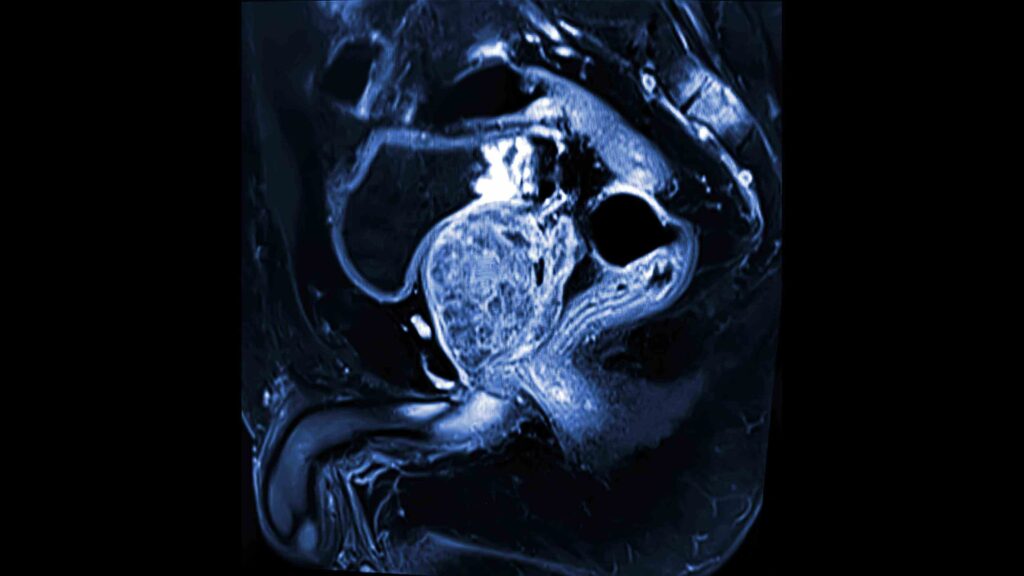
MRI scan of a prostate
The following will overview various diagnostic methods for detecting prostate cancer.
This is the most frequently used and simplest method to look for signs of prostate cancer. The doctor will slowly insert their gloved finger into the patient’s rectum to feel for lumps, prostate enlargement or other changes. If they detect something abnormal, the doctor will likely recommend additional testing to rule out or confirm cancer or other prostate conditions.
Also called prostate-specific antigen (PSA) tests, this diagnostic method measures the amount of PSA in the blood. If the results are higher than baseline expectations, it can be a sign of prostate cancer and additional diagnostic steps may be taken. However, there are multiple possible causes of elevated PSA levels besides cancer. These can include natural aging, an enlarged prostate, inflammation or infection and other benign conditions. PSA levels are also used for patients already diagnosed with prostate cancer as a way to monitor treatment efficacy.
Ultrasounds work through the use of high-frequency sound waves. A handheld device called a transducer is used to create these sound waves, which echo through the body’s internal structures and bounce back to create images that can be used for study and evaluation. When trying to detect prostate cancer, the transducer is inserted a short distance into the patient’s rectum, as the prostate is located nearby. This is called a transrectal ultrasound. The scan can help detect abnormalities, growths, nodules, enlargement and other possible signs of cancer.
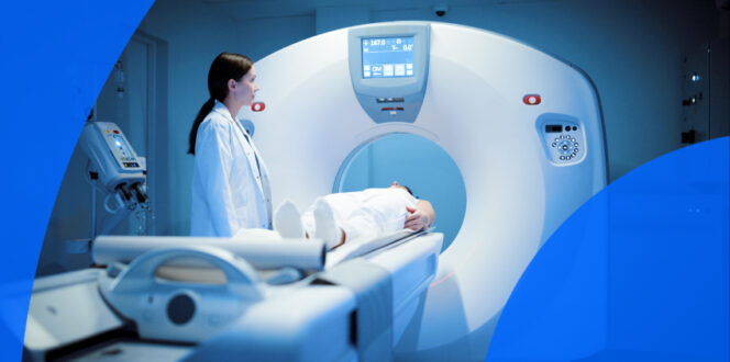
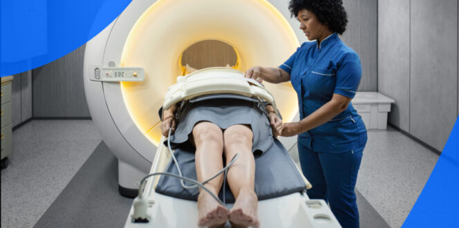
Magnetic resonance imaging (MRI) scans use strong magnets and radio waves to capture images inside the body. They can be highly detailed, allowing physicians to get a clearer look at possible findings or irregularities. During the scan, the patient will lie still in a tube-like MRI machine. They will be given hearing protection during the appointment, as the machine can be loud, producing various hums and clicks while it performs the scan. For patients with claustrophobia or anxiety, doctors may be able to prescribe calming medications if discussed in advance. There are also some open MRI machines, where the sides of the tube are open, which some patients find helpful.
A biopsy is when a small tissue sample, fluid or cells are removed from the prostate for study in a laboratory. Patients may receive a transrectal biopsy, in which a hollow needle is inserted through the rectum and into the prostate, or a transperineal biopsy, where the needle is inserted through the area between the anus and scrotum and into the prostate. These biopsies are often guided by the simultaneous use of ultrasound, helping doctors access the correct locations.
Though it can cause discomfort, a numbing agent or medication is often used during the procedure. The needle also takes samples with great speed, further minimizing discomfort. The entire appointment often takes around ten minutes and can be done in a doctor’s office rather than a hospital or specialized clinic.
The biopsy sample will then be sent to a pathologist, who will evaluate it under a microscope for cancer cells. From there, they can determine the grade and Gleason score of the cancer, if applicable. This will be discussed in the next section.
There are multiple types of cell patterns that can be detected in prostate cancer cells. These patterns can help indicate how aggressive the cancer is, which will guide treatment options and next steps. The Gleason Grading System uses these patterns to create a final score, obtained by combining the scores for the most aggressive cell type detected and the most dominant cell type detected. These numbers are then interpreted as follows:
Prostate cancer grading and staging can be complex in terms of categories and classifications. Much depends on factors like Gleason scores, PSA levels and imaging tests, which will be discussed next. Once a thorough analysis of these factors has been completed, the doctor may assign one of the following stages:
One of the most significant aspects of cancer staging, aside from PSA tests and the pathology report, is medical imaging, which helps determine if the cancer has spread and where. Below is an overview of the imaging methods used to help stage the disease.
MRI scans are useful for both the diagnosis and staging of prostate cancer. Magnetic resonance imaging can detect abnormalities and growths, as well as determine whether the cancer has spread to other regions of the body. One strength of MRI scans is the option to use a contrast agent, which is a dye injected into the patient’s arm. It provides better differentiation and detail in the imaging, allowing the radiologist to get a clearer view of any possible cancer and whether it has spread.
Computed tomography (CT) scans use low-dose X-ray technology to create internal images of the patient’s body. While standard X-rays typically produce only two to four images, CT scans generate many images, sometimes called slices. These slices are then combined to form a detailed image that can help detect prostate cancer and show whether it has spread to other areas of the body. Similar to MRIs, CT scans can be used with a contrast dye to provide improved detail and visibility.
Positron emission tomography (PET) scans are a particularly useful imaging method for detecting cancer. A mildly radioactive sugar, called a radiotracer, is injected into the patient and given time to be absorbed by their tissues. A special camera is then used to evaluate how various tissues and organs have absorbed the radiotracer.
Cancer cells tend to absorb more of the radiotracer, which can be detected in the scan. This is a highly effective way to track where the prostate cancer is and whether it has spread. Often, CT scans and PET scans can be performed at the same time, as some imaging equipment can carry out both methods with a single machine.
Much like PET scans, a bone scan uses radioactive tracers to track how the body absorbs the material. These types of imaging tests are called nuclear scans due to the use of radioactive substances in the process. They are considered safe and highly effective for assessing the spread of cancer to other areas of the body. In the case of bone scans, the focus is on determining whether prostate cancer has metastasized to the bones, which can occur in later stages of the disease.
Turnaround times for prostate cancer imaging results can vary depending on the facility and your doctor’s availability. Often, patients wait a week or more and receive their results during a follow-up appointment. With PocketHealth, you don’t have to wait as long—your results are available securely as soon as they’re uploaded, allowing you to review them before your follow-up visit.
When you do gain access to your results, it’s common to find medical terminology somewhat confusing, but Report Reader helps you to better understand your report by offering clear, straightforward definitions for medical terms—simply tap or click on any underlined words to reveal their meaning.
Here are some common questions regarding prostate cancer.
There are a variety of possible treatments for prostate cancer, including:
There are certain risk factors that may increase the likelihood of developing prostate cancer, though many people with these factors will never experience the disease. However, it can be helpful to be aware of them so individuals can better understand their personal risk. These include:
There is a very high five-year survival rate for prostate cancer cases that haven’t metastasized, currently around 99%. While cases that have spread to distant regions of the body have higher mortality rates, the majority of prostate cancer cases are detected earlier. It is also common for prostate cancer to grow very slowly or not at all, which contributes to a positive outlook for many patients, especially when the disease is caught early.
The US Preventive Services Task Force recommends the following prostate cancer screening guidelines:
Getting a prostate cancer diagnosis can feel overwhelming, but remember that you’re not alone. Thanks to advances in diagnosis and treatment, many patients experience good outcomes and an improved quality of life during and after treatment. Still, finding support and reliable resources can make a meaningful difference. Your doctor will likely have a list of local support options for you. In the meantime, here are some online resources that may also be helpful:
PocketHealth makes it simple to keep track of your scans and other medical results. All of your imaging records are in one secure location and can be accessed online anytime. If needed, reports can also be easily shared with other physicians in your care team. When used in conjunction with your medical provider’s professional advice, it is a powerful tool for organizing and understanding your imaging results and your health.
Another tool from PocketHealth is MyCare Navigator, which provides personalized insights to your health and identifies any recommended follow-up steps. This feature can also generate individualized questions to ask your doctor based on the findings in your report, ensuring you make the most of your consultation.
Published: June 4, 2025
Trusted by more than 900+ hospitals and clinics.