Thyroid Cancer Diagnosis: An Overview of Detection Methods
Thyroid cancer is less common than many other cancers but still affects thousands of patients in the United States. This disease occurs in the butterfly-shaped endocrine gland that plays an important role in regulating the body’s hormones and basic functions such as metabolism. This article will give an overview of the thyroid, discuss how thyroid cancer develops and highlight the various methods physicians use to reach a diagnosis.
What is thyroid cancer?
The thyroid gland is located in the lower front of the neck near the clavicle. Its role is to produce specific thyroid hormones: thyroxine or tetraiodothyronine (T4) and triiodothyronine (T3). These hormones are secreted and used throughout the body, affecting functions such as metabolism, menstruation, heart rate and body temperature.
Thyroid cancer develops when cells in the thyroid grow and spread out of control, affecting the function of the gland and sometimes spreading to other parts of the body such as the bones and other organs. There are multiple types of thyroid cancer to be aware of:
- Papillary thyroid cancer: This is by far the most commonly diagnosed type of thyroid cancer, making up 70 to 80 percent of cases. It can develop at any age and is usually very slow-growing. Prognosis is typically excellent, even if the cancer has spread to the lymph nodes. There are also subtypes of papillary thyroid cancer. The follicular variant is the most common, but others include hobnail, diffuse sclerosing, clear cell, trabecular, tall cell, columnar and insular. These variants may require more assertive treatment depending on the type.
- Follicular thyroid cancer: This type makes up roughly 10 to 15 percent of thyroid cancer cases. It is not the same as the follicular subtype of papillary cancer, but it also tends to have a fair prognosis. Follicular thyroid cancer is more frequently found in regions where people have inadequate dietary iodine.
- Oncocytic carcinoma: This rare cancer type makes up only about 3 percent of cases. It can be more challenging to detect and treat compared to other types. It was previously known as Hurthle cell cancer.
- Anaplastic (undifferentiated) carcinoma: Even rarer than oncocytic carcinoma, anaplastic carcinoma is found in about 2 percent of thyroid cancer cases. It can spread more quickly and be more challenging to treat compared to other thyroid cancer types.
- Medullary thyroid cancer: MTC makes up approximately 5 percent of thyroid cancer cases. It may be more difficult to treat. There are two types of MTC: sporadic and familial. Sporadic MTC accounts for the majority of cases and most often develops in older patients. Familial MTC is inherited and can develop in childhood or early adulthood.
What are the symptoms of thyroid cancer?
Thyroid cancer tends to be less symptomatic than other forms of cancer, and some patients may not experience any obvious signs at all. However, possible symptoms may include:
- A lump in the neck: A thyroid lump, nodule or swelling may be visible or may be felt by a healthcare provider during an exam.
- Difficulty swallowing: Some patients may experience trouble swallowing.
- Neck pain: Pain in the front of the neck that may also travel up to the ears.
- Chronic cough: A persistent cough that is not related to other illnesses.
- Breathing difficulties: Issues with breathing that are unrelated to respiratory conditions.
- Vocal changes: A hoarse voice or other changes in vocal tone or strength.
The presence of these symptoms does not necessarily indicate thyroid cancer. Many benign conditions can cause similar signs. Even a lump in the thyroid is often a noncancerous nodule. Still, it is recommended to speak with your physician about any symptoms or concerns so a personalized evaluation can be performed.
Thyroid cancer diagnosis methods
The definitive gold standard for confirming a thyroid cancer diagnosis is usually a biopsy. However, various blood tests and imaging studies are often performed first to help confirm or rule out any suspicion of cancer. Below is a brief overview of the different diagnostic methods.
Physical exam
A physician may feel a nodule or lump in the patient’s neck during a physical exam, especially if the patient is experiencing symptoms. Some providers check for thyroid lumps as part of routine examinations.
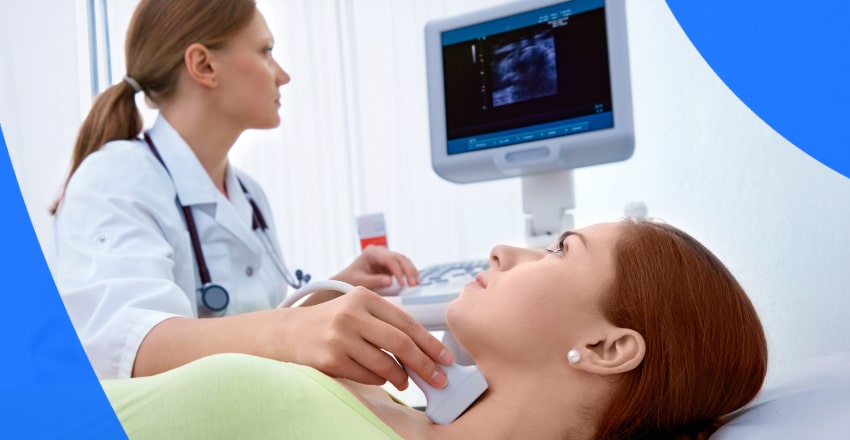
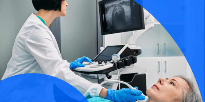
Ultrasounds
Ultrasound scans use high-frequency sound waves to create internal images of the body. These sound waves are produced by a handheld device called a transducer, which is slowly moved over the area being scanned. In this case, the area is the patient’s neck. The waves echo off internal structures and tissues, creating real-time images that the technician can capture and save for later analysis.
Ultrasounds are particularly helpful in diagnosing thyroid cancer because it can differentiate between a fluid-filled and a solid nodule or growth. Fluid-filled nodules tend to be benign, while solid nodules have a higher likelihood of being cancerous. Ultrasounds can also assess nearby lymph nodes, as enlargement may indicate the cancer has spread.
Lastly, ultrasounds are commonly used to help guide biopsy needles into the correct nodule or area being sampled. Its real-time imaging and accuracy make it a valuable tool for this purpose.
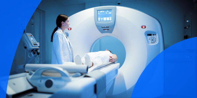
CT scan
Computed tomography scans use X-ray technology to create highly detailed cross-sectional images of the body. Traditional X-rays typically capture only 2 to 4 images for analysis. CT scans, however, take several images and combine these numerous “slices” into a detailed view that can reveal nodules, abnormalities and signs that the cancer has spread to other areas of the body.
Often, a contrast dye is used during the CT scan, injected into the patient’s arm. This dye helps differentiate the areas being scanned, improving visibility and detail.
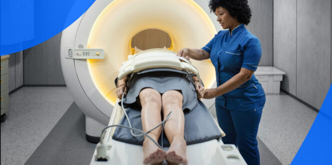
MRI Scan
Magnetic resonance imaging uses strong magnets and radio waves to produce highly detailed internal images for study. In the case of thyroid cancer, it can show the gland and adjacent lymph nodes, detailing factors such as size, tumors and abnormalities. It can also help detect if cancer cells have spread to other areas of the body, though PET scans and other imaging methods are more commonly used for this purpose. Similar to CT scans, MRIs can also be performed with a contrast agent.
PET scan
Positron emission tomography (PET) scans are extremely valuable in detecting cancer throughout the body. A mildly radioactive radiotracer, often fluorodeoxyglucose (FDG), is injected into the patient before the scan. After the tracer has had time to absorb into the body, the patient undergoes imaging in the PET scanner. A special camera tracks the radiotracer, evaluating how tissues and cells absorb the substance. Cancer cells tend to absorb more of the radiotracer than normal cells due to their higher metabolic rate, which helps doctors identify affected areas and determine whether the cancer has spread.
Often, CT scans and PET scans can be performed at the same time using the same equipment, as some imaging machines are designed to do both. PET scans are particularly useful for detecting recurrent thyroid cancer, determining whether it has metastasized to other parts of the body and evaluating how effectively treatments are working.
Radioiodine scan
Also called a thyroid scan, a radioiodine scan is an imaging method in nuclear medicine that works similarly to a PET scan. Instead of a radioactive sugar such as FDG, a thyroid scan uses a radioactive iodine tracer. This is often taken in pill form or injected and given time to be absorbed by the body. During the scan, a gamma camera detects and measures the gamma radiation emitted by the tracer, capturing images of the affected areas.
Nodules or regions that absorb more radiation than expected are called hot nodules, which are often not cancerous. Those that absorb less radiation are referred to as cold nodules. Cold nodules can sometimes indicate cancer, although benign nodules can also appear this way. A thyroid scan by itself is not used to diagnose thyroid cancer, since cold nodules can have non-cancerous causes. However, it can help rule out certain conditions or determine whether additional testing, such as a biopsy, is needed.
Blood tests
There are multiple blood tests that may help assess thyroid function. Because it is not uncommon for patients with thyroid cancer to have normal blood test results, these tests are usually used to evaluate for benign thyroid conditions, such as hypothyroidism. They may help indicate that a nodule or finding is non-cancerous or determine whether additional testing, such as imaging, is needed. Thyroid blood tests commonly assess levels of:
- Thyroid-stimulating hormone (TSH): TSH is produced by the pituitary gland, not the thyroid. It helps regulate other thyroid hormones. If the thyroid is producing too few hormones, the TSH level may be higher than expected. This test helps evaluate general thyroid function.
- T3 and T4: These thyroid hormones are produced by the thyroid gland and help control metabolism.
- Calcitonin: This hormone is involved in the body’s use of calcium and is produced by C cells in the thyroid. These cells can mutate into medullary thyroid cancer, so abnormal calcitonin levels may point to this condition.
- Carcinoembryonic antigen (CEA): This protein may be elevated in patients with medullary thyroid cancer.
Biopsy
Thyroid nodules and tumors are often benign, which is why a biopsy is needed to definitively diagnose thyroid cancer. A small tissue sample is taken from the nodule and sent for laboratory analysis to check for the presence of cancer cells. Typically, an ultrasound or other imaging is performed first to help the physician determine whether a biopsy is necessary. Biopsy types include:
- Fine needle aspiration (FNA): FNA is one of the most common biopsy types and is often performed in a clinic or doctor’s office. A hollow, thin needle is inserted through the skin into the nodule, often with real-time guidance from ultrasound. A small sample is removed, and the process may be repeated as needed, including in the lymph nodes if they appear to be affected. The procedure is quick, minimally invasive and considered very safe.
- Core needle biopsy: This is similar to fine needle aspiration but uses a larger needle to obtain a more substantial tissue sample. It often requires the procedure to be performed by someone with specialized training, as it is more invasive than FNA.
- Surgical biopsies: If results from previous biopsies are unclear, a larger sample may be needed. This can be obtained during a surgical biopsy, typically performed under general anesthesia. One example is a lobectomy, which involves removing the lobe of the thyroid that contains the nodule or cancerous cells. A lobectomy is also a common treatment for thyroid cancer.
Getting your results
Turnaround times for thyroid cancer imaging results can vary depending on the facility and your doctor’s availability. Often, patients wait a week or more and receive their results during a follow-up appointment. With PocketHealth, you don’t have to wait as long—your results are securely accessible as soon as the imaging or report are available, allowing you to review them before your follow-up visit.
When you do gain access to your results, it’s common to find medical terminology somewhat confusing, but Report Reader helps you to better understand your report by offering clear, straightforward definitions for medical terms—simply tap or click on any underlined words to reveal their meaning. This is paired with illustrations and highlights of anatomy in your imaging to help you better understand your results and prepare for follow-up appointments.
Frequently asked questions
Here are some common questions regarding thyroid cancer.
What is the outlook for thyroid cancer?
Outlook and prognosis for thyroid cancer vary depending on whether the cancer has spread to distant parts of the body (a process called metastasis), the type of thyroid cancer and the patient’s overall health. Many common variants of thyroid cancer, such as papillary or follicular, have high survival rates, especially if the cancer is localized and hasn’t spread. Regarding your own personal case, it is recommended to discuss your medical history with your doctor so they can provide a more accurate assessment of your individual health. Overall, thyroid cancer tends to have very high rates of successful treatment.
What causes thyroid cancer?
There is no single known cause of thyroid cancer. Cells may mutate for no obvious reason, or the mutations may be inherited and run in a patient’s family. In some cases, radiation exposure during childhood, such as exposure during treatment for a prior cancer, can increase the risk of developing thyroid cancer later in life.
How is thyroid cancer treated?
There are multiple possible methods for treating thyroid cancer, and many are used in combination with one another. Some common treatments include:
- Surgery: Part of the affected thyroid may be surgically removed, or in some cases, the entire thyroid gland. This is one of the most common treatments. If the cancer has spread to nearby lymph nodes, those may be removed as well.
- Radiation therapy: High-energy rays, such as X-rays, are used to destroy cancer cells and prevent them from spreading.
- Radioactive iodine therapy (RAI): RAI is usually administered orally. The radioactive iodine collects in thyroid tissue, including cancerous cells, and destroys it. Other types of tissue remain unaffected, as only thyroid cells absorb iodine.
- Chemotherapy: Special drugs that help kill cancer cells and keep them from growing. This is more commonly used for aggressive forms of thyroid cancer.
- Hormone therapy: If cancer cells are feeding off of specific hormones, medications to block or stop these hormones may also help kill the cancer and keep it from spreading.
- Targeted therapy: Medications that target specific aspects of thyroid cancer cells. One example is a protein inhibitor that blocks certain proteins used as fuel by cancer.
- Immunotherapy: This new and still developing treatment involves bolstering the patient’s immune system to fight the cancer, and helping it better recognize cancer cells as threats.
What does the thyroid do?
The thyroid gland produces triiodothyronine (T3), thyroxine or tetraiodothyronine (T4) and calcitonin, the last of which helps regulate calcium levels. These hormones help regulate body temperature and heart rate, support growth and brain development during childhood, activate the nervous system and play a role in various metabolic functions.
Getting support
Getting a thyroid cancer diagnosis can be an overwhelming experience but it’s important to know you’re not alone. Your doctor will likely have local resources and support for you to take advantage of, and there are numerous online resources you may find helpful as well:
Take control of your health journey
PocketHealth makes it simple to keep track of your thyroid scans. All of your vital imaging is in one secure location and can be accessed online anytime. Imaging and reports can also be easily shared with other physicians in your care team, if needed. Additionally, it makes it easy to track health changes over time, such as with repeat imaging. When used in conjunction with your medical provider’s professional advice, it is a powerful tool for organizing and understanding your imaging results and your health.
PocketHealth MyCare Navigator gives personalized insights into your health and identifies any recommended follow-up steps. This functionality can also generate individualized questions to ask your doctor based on the findings in your report, ensuring you make the most of your consultation. A thyroid cancer diagnosis may feel overwhelming, but working closely with your providers can give you the best chance of managing your condition and protecting your health.
Published: July 8, 2025
Trusted by more than 800+ hospitals and clinics.