Thyroid eye disease, also called TED, is an autoimmune condition that can affect some patients with thyroid disorders, including hyperthyroidism and, in rare cases, those with normal or low thyroid levels. These conditions can sometimes impact the tissues and areas around the eyes, leading to pain and inflammation that may become problematic.
This article will provide a basic understanding of the thyroid eye disease, outline its symptoms and discuss the different diagnostic methods used to assess patients for TED.
The thyroid gland is located at the base of the front of the neck. Shaped like a butterfly, its purpose is to produce thyroid hormones, which are used throughout the body to regulate metabolism, heart rate, blood pressure, bone health, growth and development and other functions.
There are various autoimmune disorders where the immune system mistakenly attacks healthy tissues instead of protecting them. In the case of TED, the immune system targets the thyroid gland, which can lead to inflammation in the tissues around the eyes. This happens because the proteins in the eye tissues are similar to those in the thyroid, causing the immune system to mistakenly attack both areas.
One form of this autoimmune condition is called Graves’ disease. Since thyroid eye disease is a common response to having Graves’ disease, the two conditions are often termed interchangeably, even at times, within the medical community. However, it’s important to remember that some patients may experience TED without actually having Graves’ disease, as there are multiple thyroid conditions that may cause TED. Other terms for thyroid eye disease include:
For many patients, TED occurs in specific phases. The initial or “active” phase, which may last from six months to two or more years, is when inflammation tends to be most pronounced. Symptoms may fluctuate from minor to severe, coming and going during this time. In most patients, this stage will eventually subside, and the eye changes will stabilize.
For those who experience particularly severe changes during this first phase, there may be permanent damage or structural changes, which could require treatment or surgery to improve function. Other patients, however, may see their eyes return to a normal state. Fortunately, after this phase ends, it’s unusual for eye changes to occur again, meaning many patients have good treatment options and outlooks.
Symptoms of thyroid eye disease may vary from patient to patient. Many individuals will only experience mild symptoms, though in some cases, they can range to severe. Here are some possible symptoms of TED:
It is important to remember that these symptoms may be caused by numerous medical conditions besides thyroid eye disease. If you have concerns, consider discussing them with your doctor so they can make any assessments and recommendations for your individual health.
Some of the possible factors that increase a patient’s risk for developing TED include:
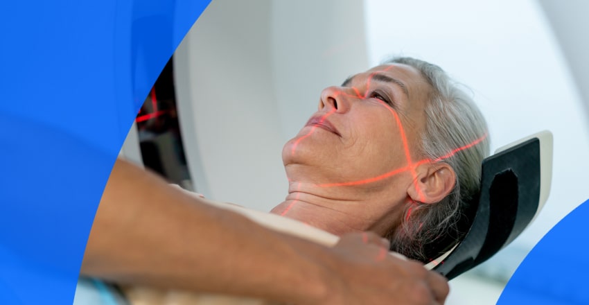
MRI of the head and eyes
There are multiple methods to diagnose thyroid eye disease. Here is a brief overview of the most common types.
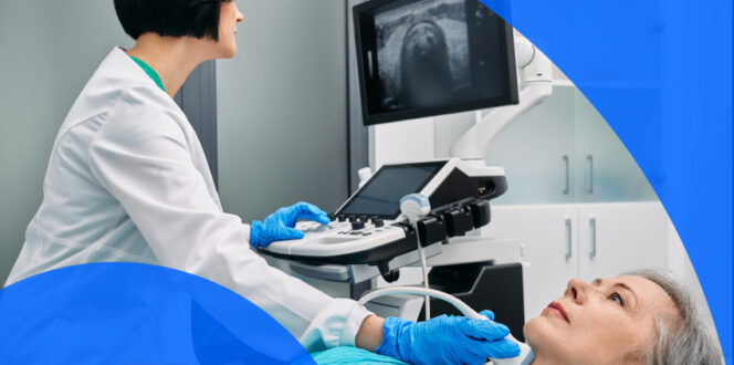
Because TED can cause visual symptoms, such as double vision, an important step in diagnosis is assessing the patient’s field of vision and other eyesight functions. If the patient is experiencing any vision loss, it’s important they inform their doctor immediately, as there are often medications and treatments to help manage this.
Another component of the diagnosis is a physical eye exam, where the doctor will take measurements of any eye protrusion or bulging, note any eyelid retraction, inflammation or other changes in the eye area.
There are a variety of blood tests that can help diagnose TED, including checking for too much thyroid hormone (or too little), or other indicators of an underlying thyroid disorder, such as Graves’ disease. These blood tests can also be used throughout treatment to monitor how well the condition is being managed.
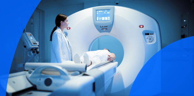
Computed tomography (CT) scans are one of the imaging methods used to assess thyroid eye disease. CT scans use low-dose radiation X-rays to capture detailed images of internal structures. Multiple X-ray images are taken and then merged together to create a more in-depth view of the area being evaluated. In this case, the eyes and surrounding tissues would be scanned to look for inflammation, swelling, optic nerve damage and other structural changes.
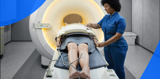
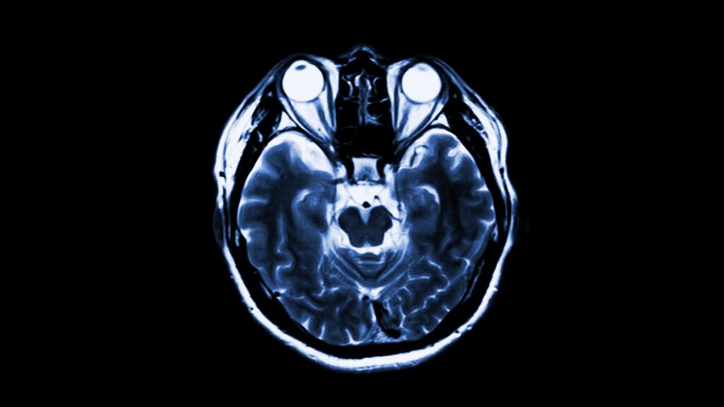
Magnetic resonance imaging (MRI) is another imaging method used to diagnose TED. MRIs use radio waves and strong magnets to align the protons in the body’s hydrogen atoms. The MRI machine then sends a pulse of energy, temporarily changing their alignment. When these protons return to their previous position, they release energy, which the MRI machine picks up and uses to create detailed internal images. Similar to a CT scan, the MRI is used to look for changes and inflammation in the eye muscles, surrounding tissues and other optical anatomy.
There are a variety of helpful treatments for thyroid eye disease. Many cases of this condition are mild, so treatments may be fairly simple. For more severe cases, there are still numerous options to help manage symptoms. Here are some common methods:
Turnaround times for imaging results can vary widely depending on the facility and your doctor’s availability. Often, patients wait a week or more and receive their results during a follow-up appointment. With PocketHealth, you don’t have to wait as long—your results are securely accessible as soon as the report is available, allowing you to review them often before your follow-up visit.
Your imaging results are interpreted by a radiologist—an expert in medical imaging—who carefully reviews your scans and provides a detailed report of any findings. This report is then sent to your referring doctor, who, with a deeper understanding of your medical history, can offer further insights and recommendations.
To better understand your imaging reports, Report Reader provides clear, straightforward definitions for medical terms. Simply tap or click on any underlined words to reveal their meaning. This is paired with illustrations and highlights of anatomy in your imaging to help you better understand your results and prepare for follow-up appointments.
Here are some common questions regarding thyroid eye disease.
Everyone is different, and symptoms may vary from patient to patient. However, many people find that initial signs of the disease include eyelid retraction, bulging eyes, aching behind the eyes or sensitivity to light.
For severe cases that aren’t medically controlled, thyroid eye disease can cause long-term complications such as permanent vision loss or double vision, eyelid changes and other lasting effects. Many of these complications have treatment options to help manage or reduce symptoms, and getting treatment early can often prevent complications from becoming permanent.
You may come across multiple resources that use the terms Graves’ disease and thyroid eye disease interchangeably. While many patients with Graves’ disease also have TED, they are technically separate conditions. Both are autoimmune diseases that affect the thyroid, but it’s possible to have TED without Graves’ disease, and it’s possible to have Graves’ disease without TED.
CT and MRI scans are highly detailed and accurate at detecting inflammation, swelling and other abnormalities in the tissues around the eyes. However, thyroid eye disease may involve multiple diagnostic components to confirm an official diagnosis. In addition to medical imaging, blood tests, eye exams and assessments of thyroid function can help ensure there are no other causes for eye symptoms.
PocketHealth makes it simple to keep track of your eye imaging. All of your vital imaging is in one secure location and can be accessed online anytime. Reports can also be easily shared with other physicians in your care team, if needed. Additionally, it makes it easy to track health changes over time, such as with repeat imaging. When used in conjunction with your medical provider’s professional advice, it is a powerful tool for organizing and understanding your imaging results and your health.
PocketHealth MyCare Navigator gives personalized insights into your health and identifies any recommended follow-up steps. This feature can also generate individualized questions to ask your doctor based on the findings in your report, ensuring you make the most of your consultation.
Published: May 16, 2025
Trusted by more than 900+ hospitals and clinics.