
Mammogram screening is a valuable tool to assess your breast health, and regularly scheduled mammograms help your doctor catch breast abnormalities that could be cancerous. Finding breast cancer early is critical to the effective treatment of the disease, and regular mammogram screenings can help alert your doctor to new changes in breast tissue.
New draft 2023 mammogram guidelines issued by the U.S. Preventive Services Task Force (USPSTF) suggest that a woman at average risk of breast cancer should receive a mammogram every two years beginning at 40 years old. Recent research indicates that regular mammograms between 40 and 49 years old reduce breast cancer mortality risk by 12-29%.
Some women may be at greater risk and may have mammogram screenings more often. Your doctor may send you for a mammogram as young as 30 years old if you are a carrier of the BRCA1 or BRCA 2 gene or have had radiation to your chest since both factors increase your risk of developing breast cancer.
If it’s time for your mammogram screening you may have questions about the appointment, including:
Understanding how mammograms work and how regular screening benefits you, lets you stay on top of your health and have more informed discussions with your physician about the results.
A mammogram is an X-ray that allows your healthcare team to assess the tissue of your breast for irregularities, including lumps. Mammograms help your doctor investigate any abnormalities you may feel while doing a breast self-exam, but they can also catch lumps too small for you to feel.
Mammograms use X-ray technology to examine breast tissue. While very small amounts of radiation are released during a mammogram, the benefits of catching breast cancer early outweigh the radiation exposure for most. If you have concerns about the impact of the radiation from a mammogram, speak with your doctor to better understand the risks of the procedure.
There are 3 different types of mammograms:
After your mammogram screening test, the mammography technician will send the images of your screening to a radiologist. A radiologist is a doctor who specializes in diagnosing and treating injuries and diseases using medical imaging. Once a radiologist has assessed your mammogram images, they will send the images and a report to your doctor, who will schedule an appointment to discuss any results. PocketHealth enables patients to access their mammogram results as soon as the radiologist has completed their report. Access your records here.
Women with average breast density should begin getting regular mammograms at age 40. New U.S. breast cancer screening guidelines for 2023 state that receiving regular mammograms from age 40 onwards can reduce the risk of serious breast cancer outcomes and mortality.
Breast tissue density is an important factor when considering breast cancer risk and screening. A mammogram can show the radiologist how much dense fibroglandular tissue is in your breasts. Women with very dense breasts, containing a lot of fibroglandular tissue, have an increased risk of developing breast cancer but should still receive regular mammograms. Dense breast tissue can make mammogram screening less effective for young women, so speak to your doctor if you’re concerned you may fall into this category.
Whether or not you have any signs or symptoms of breast disease, the USPSTF draft recommends a mammogram screening every two years for all women at average risk of breast cancer between the ages of 40 and 74 years of age.
Reasons you should have regular mammogram screenings include:
For the technologist to get the clearest images of your breast tissue, the breast must be compressed, which can cause some discomfort. Avoiding a mammogram in the 10-14 days before your menstrual period can help reduce discomfort and if you have a breast condition that makes your breasts sensitive or tender, speak to your doctor about taking pain medication before your appointment.
Mammogram imaging allows the radiologist to see very small abnormalities in breast tissue, even those too small to feel with a physical exam, and is very effective at screening for breast cancer. Mammograms allow malignant abnormalities to be detected early, increasing the effectiveness of cancer treatment. Research in 2020 shows that mammogram detection of breast cancer early on improves recovery rates and reduces death from breast cancer by 41% over the ten years following detection.
Although regular mammogram screenings can mean cancer is caught early, making treatment more effective, mammograms also have limitations. Some cancers cannot be detected on a mammogram because of the location of the cancer or the density of breast tissue. Mammograms also release a small amount of radiation during imaging, but this low amount does not outweigh the benefits of regular mammograms, according to the National Cancer Institute. If you are concerned about radiation from mammogram screenings, talk to your doctor about the risk to you.
Mammograms can give false-positive or false-negative results, meaning the images do not accurately represent what’s happening in your body. With a false-negative result, a mammogram indicates that there are no concerning irregularities in your breast tissue— when there are. On the flip side, a false-positive result indicates there is an area of concern where there isn’t one.
A 10-year research study that false-positive results happen about 12% of the time, but that only 4% of those mammograms indicate a “positive” result in a diagnosis of cancer. The National Cancer Institute states that mammograms give false-negative results, missing cancer that’s present at the time of screening, about 20% of the time.
False-positive and false-negative mammogram results can cause a lot of stress to the patient and lead to unnecessary testing or, in some cases, can cause cancerous results to be missed.
A mammogram is an integral part of breast cancer screening and requires only a short appointment, but there are some steps you can take to prepare in advance:
The imaging clinic or department will provide wipes to clean up after your screening, but you may wish to pack deodorant or antiperspirant to bring with you to wear after the appointment. Also, if you are concerned about pain during your appointment, ask your doctor if over-the-counter medication can help reduce discomfort.
During your mammogram appointment, the technologist will go over the procedure with you and outline each step. If you have any questions about the details of the process or what to expect, you can ask for clarification before the screening begins.
During your mammogram appointment, you can also ask:
After your mammogram appointment, your doctor will schedule a follow-up call or in-person appointment to discuss your results. At this appointment, you can ask any questions about your results, such as:
Your imaging results are interpreted by a radiologist—an expert in medical imaging—who carefully reviews your scans and provides a detailed report of any findings. This report is then sent to your referring physician, who, with a deeper understanding of your medical history and past exams, can offer further insights and recommendations on next steps in a follow-up appointment.
Turnaround times for imaging results can vary widely depending on the facility and your doctor’s availability. Often, patients wait a week or more and receive their results during a follow-up appointment. With PocketHealth, your imaging results are securely accessible as soon as they’re approved for release by the hospital or imaging clinic. This allows you the opportunity to review your results and prepare questions ahead of your follow-up visit.
To help you understand your mammogram results, PocketHealth provides clear definitions and illustrations for complex medical terms—plus an in-depth explanation of your full imaging report. This is paired with highlights of key anatomy in your imaging to help you better comprehend what you’re looking at.
“I recently had mammograms taken and was to see my doctor in five weeks to hear the results. PocketHealth gave me the results as soon as they were ready. While my results were clear, PocketHealth gave me peace of mind because, without it, I would have worried for weeks about my results.”
– Mammogram Screening Patient
Mammograms can show different areas of irregularities within your breast tissue, not only lumps or masses. Both normal and abnormal mammogram images can show other parts of your breast tissue that aren’t cancerous but may indicate a different health concern or may be completely benign, including cysts, fibroadenomas or calcifications. Here’s a list of common irregularities in breast tissue found by mammograms:
Normal, abnormal and cancerous mammogram images will show some differences, but all mammograms will have a black background and show the image of the breast in variations of gray and white. In general, denser tissue appears white and less-dense tissue, including fat, appears gray.
Therefore, a normal mammogram image of healthy breast tissue will show primarily dark areas interspersed with some lighter or cloudy-looking areas. Abnormal images may show thicker, light areas of more dense tissue, or smaller light areas of dense tissue, while a mammogram image indicating breast cancer will likely show one or more clearly defined white, opaque masses within the breast tissue.
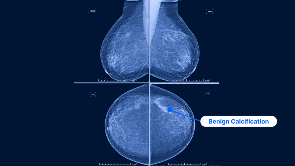
A mammogram image showing two standard views: the MLO view (i.e. taken from the side) is on the top and the CC view (i.e., taken from above) is on the bottom. The CC view of the left breast more clearly shows a benign calcification with no evidence of malignancy, falling into BI-RADS category 2.
Created by the American College of Radiology, BI-RADS is used to report the results of every mammogram. The system accounts for the results of the mammogram and recommends follow-up tests, if needed, and indicates the degree of the risk that an abnormality may be cancerous.
BI-RADS works by organizing the results of each mammogram into 7 categories based on cancer risk. Each category is given a number, with lower numbers indicating no risk or benign changes and higher numbers indicating areas of breast cancer concern. A BI-RADS category of “0” indicates an incomplete scan, so additional testing or a repeat mammogram is required.
Depending on the results of your mammogram, your doctor may send you for other breast cancer screenings, including a breast ultrasound, a breast MRI or molecular breast imaging. These tests can be useful in determining whether a lump is a hard mass or a fluid-filled cyst. If cancer has been detected, these additional screenings can indicate how advanced the disease is. In some cases, different breast cancer screenings will be used in conjunction with each other to ensure no cancer has been missed.
While breast cancer continues to be one of the most common forms of cancer among women, regular mammogram scans can significantly reduce your risk of serious breast cancer complications. If you’ve missed or delayed a regular mammogram screening, it’s never too late to start or to catch up—early detection of breast cancer can improve treatment outcomes.
If you’re approaching 40, speak with your doctor about regular mammogram screenings and discuss the benefits and limitations of the technology, as well as your cancer risk.
If you’re located in Ontario, you have the option to get your breast cancer risk score and determine your eligibility for Ontario Breast Screening Program (OBSP), as part of PocketHealth’s suite of breast health tools. If you’re eligible for OBSP, you can even book your next mammogram directly through PocketHealth.
With PocketHealth, it’s simple to keep track of your mammogram imaging results. All of your images and reports are permanently available in one secure location and can be accessed online—anytime, anywhere. Reports can also be easily shared with other members of your care team, if needed. When used in conjunction with your medical provider’s professional advice, it is a powerful tool to better understand your imaging results.
PocketHealth also provides personalized health insights based on the findings in your report to help you stay on top of any next steps. This includes clearly surfacing any follow-up actions found in your report and generating customized questions to ask your doctor so you can make the most of your follow-up appointment.
Learn more about how to use PocketHealth to access and share your mammogram records.
Published: August 30, 2023
Trusted by more than 900+ hospitals and clinics.