
If your doctor has recommended scheduling a bladder ultrasound, you may have questions about this imaging technique. This guide will explain its uses, the conditions it can help diagnose, and how to prepare for your appointment.
While this guide provides helpful information about bladder ultrasounds, your doctor will interpret your results officially during a follow-up appointment or phone call. If you’d like early access to your results, PocketHealth offers secure, early access to your report when it is available. This allows you to review the findings in advance and prepare any questions for your follow-up.
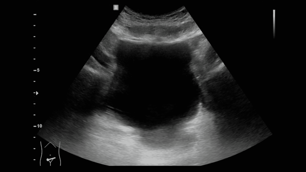
Ultrasound of the bladder
Ultrasounds are an imaging technology that uses high-frequency sound waves to create two-dimensional pictures of a patient’s internal structures. As the sound waves bounce off organs and bones, the echoes reflect back to the transmitter, allowing the images to be captured for analysis. A bladder ultrasound specifically focuses on this area, where urine is stored after being processed by the kidneys.
Bladder ultrasounds can help diagnose and evaluate a range of health conditions that affect this area. Here are some common reasons medical providers use these scans:
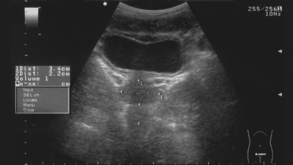
Having a full bladder is usually required for a bladder ultrasound. This improves image quality, as sound waves move faster and more densely through liquid, providing better detail. Additionally, many patients will need to have both a full and empty bladder measured. Here’s what to expect during the ultrasound appointment.
Here are some tips to help you prepare for your ultrasound appointment:
Understandably, many patients are eager to receive and go over their bladder ultrasound results. This section will discuss common questions regarding when to expect your results and who deciphers them.
A radiologist, a specialist in diagnosing and assessing conditions through medical imaging, will interpret your ultrasound images. The radiologist’s findings will then be sent to your referring physician, who may provide additional insights or diagnostic interpretations based on your overall medical history.
Depending on the radiologist’s and your referring physician’s schedules, it may take up to a week or longer to receive your results, though this can vary by facility and situation. Most patients receive their results during their follow-up appointment with their doctor.
If you’d like to view your report before your appointment, PocketHealth provides faster access, allowing you to see the results as soon as they are officially uploaded. This gives you time to review the findings and prepare for your next medical visit. MyCare Navigator is especially helpful for offering personalized insights and identifying any recommended follow-up steps. It recommends questions based on your personal report that you may want to ask your doctor, helping you make the most of your consultation.
Although your doctor will provide an official explanation of your results, accessing your report before your appointment can give you an early glimpse. It’s common to find medical terminology complex and confusing, but PocketHealth Report Reader makes it easier by offering clear, straightforward definitions for medical terms—simply tap or click on any underlined words to reveal their meaning. Meanwhile, here is a quick overview of common findings for bladder ultrasounds.
Ultrasounds can provide information on bladder and ureter anatomy and size, the thickness of the bladder wall, the bladder’s capacity and the amount of urine remaining in the bladder after urination. This is known as post-void residual volume. If these assessments are within normal ranges, you may see terms such as:
Possible abnormal bladder ultrasound findings include:
Ultrasound of the bladder and prostate
Often, an ultrasound is the first step in diagnosing bladder conditions. Sometimes, it is the only diagnostic step needed, but frequently, additional tests or scans are performed. Here are some common testing progressions for patients with bladder symptoms:
Bladder ultrasounds use high-frequency sound waves to capture images, whereas MRIs use magnetic and radio waves. Both are low-risk scans that don’t use radiation and are highly effective for evaluating internal areas. CT scans, however, use a small dose of radiation, though they are still considered to be safe and effective for patients.
Ultrasounds are ideal for dynamic imaging, meaning they can capture images during the patient’s movements if needed. Soft tissues, such as tendons, muscles, organs and connective tissue, show up well on ultrasound scans. However, bone and air (such as in the lungs) are less clear with this technique. Ultrasounds are also beneficial for other reasons, such as ease of scheduling and being less intimidating for patients who may feel claustrophobic during an enclosed MRI scan. They are typically less expensive and can accommodate patients with internal medical devices containing metal, such as pacemakers. MRIs, due to their magnetic nature, have restrictions with metal implants.
MRIs are a useful complement to ultrasounds when needed. Ultrasounds are less effective at showing structures like cartilage, bones, joints and larger areas, which are better evaluated with an MRI. MRIs can also assess soft tissues, tumors, inflammation and other conditions with great detail. Often, an ultrasound is the initial imaging step, followed by an MRI or another in-depth imaging method for further analysis. Additionally, some insurances won’t cover MRIs ( or CT scans) until an ultrasound has been performed first.
CT scans are a type of X-ray technique that expose the patient to a very small dose of radiation. While considered low-risk and safe, medical providers typically avoid having patients undergo these scans repeatedly. In terms of bladder imaging, CT scans provide excellent three-dimensional detail, helping assess a wide variety of internal structures and soft tissues. Overall, CT scans excel at spatial resolution, meaning they can clearly show the edges of structures, such as where one structure ends and another begins. MRIs, on the other hand, are better at contrasting multiple areas of the body, such as cancerous tissue compared to healthy tissue. Each has its own strengths, and neither is considered superior to the other. However, CT scans may be a better option for those who experience claustrophobia in an MRI machine or have pain issues, as CT scans are faster and more comfortable for patients.
Here are some commonly asked questions regarding bladder ultrasounds.
Bladder ultrasounds are most effective when the patient has a full bladder. However, some patients may not be able to hold their urine for the duration of the appointment. Patients should consult with their doctor or the imaging clinic for possible adjustments. Common approaches for this issue include emptying the bladder halfway to alleviate discomfort or urinating as needed while drinking more than the typical 32 ounces (946 mL) of water to compensate. Be sure to consult with your medical provider to determine their preferences.
Ultrasounds are considered a low-risk and safe imaging method. Unlike X-rays and similar scans, they do not use radiation, so they can be performed repeatedly without concerns about side effects or other issues.
Ultrasounds can be very accurate, especially for conditions that are easy to spot, such as bladder diverticula. That said, there are multiple bladder conditions that may require additional imaging or tests to confirm, which is common for most medical imaging methods. Ultrasounds serve as an excellent starting point for evaluation and diagnosis.
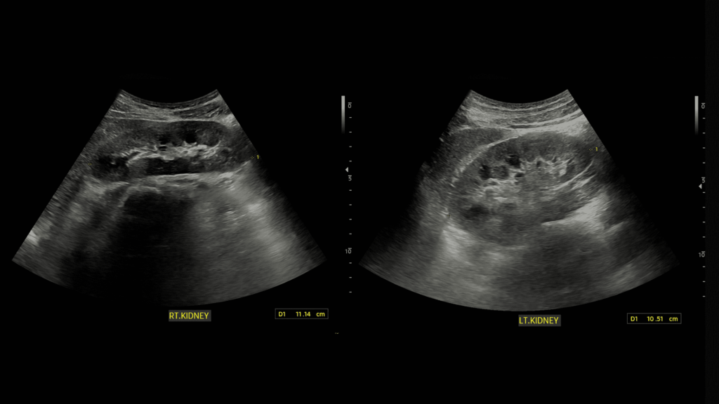
Ultrasound of the kidneys
A kidney ultrasound focuses primarily on the kidneys, but it also provides details of the ureters. Bladder ultrasounds similarly evaluate the ureters, but they focus more on the bladder itself. Because urinary conditions can affect or stem from both of these organs, it is not uncommon for patients to have a kidney and bladder ultrasound during the same appointment. The technician simply moves the transducer over the kidney and bladder areas for evaluation.
Often, patients can maintain their normal habits before their ultrasound, including eating as usual. However, some doctors may request that patients avoid caffeine and alcohol (as already mentioned) and only drink clear liquids. This is because dyes in colored liquids or their opaqueness can sometimes interfere with imaging clarity. Be sure to follow any recommendations from your provider.
PocketHealth makes it simple to keep track of your medical reports and ultrasounds. All of your medical imaging is in one secure location and can be accessed online anytime. If needed, reports can also be easily shared with other physicians in your care team and family members. When used in conjunction with your medical provider’s professional advice, it is a powerful tool for organizing and understanding your imaging results and your health.
Published: January 13, 2025
Trusted by more than 900+ hospitals and clinics.