Colorectal cancer is the fourth most common cancer in the U.S., with the average age of diagnosis being 66, though there are many exceptions. Fortunately, treatments and survival rates for colorectal cancer continue to improve, along with detection methods and screenings. When discovered early, it is highly curable, which is why there has been a strong emphasis on patients keeping up with routine screenings. This guide will walk you through what to expect when being tested for or diagnosed with colorectal cancer, including common testing methods and imaging techniques.
While this guide provides helpful information, your doctor will give the official interpretation of diagnostic testing and medical imaging results during a follow-up appointment or phone call. However, if you’d like early access to your results, PocketHealth offers secure, instant access to your medical imaging reports when they are available. This allows you to review the findings prior to your follow-up appointment and be more prepared for the discussion with your doctor.
Colorectal cancer is a broad term that can describe either colon cancer (which starts in the colon) or rectal cancer (which begins in the rectum). Because both affect the digestive system, they are often diagnosed and treated similarly, but there are distinct differences between the two, which will be described in the next section. In the meantime, here are some overall key traits of colorectal cancer:
This occurs when cells begin to form and grow out of control in the colon (the large intestine). If left undetected for a long period of time (typically about ten years), these cells often form a growth called a polyp, which can develop in the inner lining of the colon. A polyp does not necessarily mean cancer—this common condition is usually benign—but it can eventually develop into cancer or may already be cancerous when discovered. Typically, the polyp will be removed for testing or as part of treatment. Patients should remember that only 5 to 10% of polyps become cancerous.
Colon cancer typically begins in the first part of the large intestine, while rectal cancer is more commonly found in the last 12 centimeters, known as the rectum. One key difference is that the rectum isn’t as well protected as the colon, making it easier for tumors to break through the outer layers and spread. This also makes rectal cancer 20% more likely to recur in the same location, compared to just 2% for colon cancer recurrence.
Colorectal cancer may not always cause symptoms, or it may mimic the signs of more common digestive issues. This is why routine screening is so helpful for early detection, as symptoms aren’t always obvious. The following list of symptoms is worth discussing with a healthcare provider, especially if multiple symptoms are present. However, patients experiencing them should not panic, as many benign medical conditions can cause these same symptoms:
Because many colorectal cancer symptoms mimic more common conditions, it can be difficult to detect them early, especially if symptoms are absent. For this reason, the American Cancer Society recommends that individuals at average risk for colorectal cancer begin screening at age 45 and continue through age 75. This may be done using a stool-based test to check for signs of cancer (which will be discussed further in this guide) or, more commonly, through a colonoscopy.
A colonoscopy involves a physician inserting a thin, flexible tube with a camera (called an endoscope) into the patient’s anus to examine the large intestine, including the anus, rectum and colon. To prepare for this, the patient must perform bowel preparation in advance to ensure the large intestine is completely empty and unobstructed. They are also given sedation or anesthesia during the exam.
From there, the endoscope allows the provider to capture images of the large intestine for analysis and to check for abnormalities, polyps and growths. It also enables the removal of tissue samples and small polyps for testing, if needed.
Cancer screening for high-risk patients (such as those with a family or personal history of colorectal cancer) varies depending on symptoms, past treatments and other factors. They may undergo colonoscopies or other tests more frequently or at earlier ages. Otherwise, the screening recommendations for patients at average risk include:
There are multiple possible first steps to diagnosing or ruling out colorectal cancer. The following is a brief overview of these methods.
Also called a DRE, this exam involves the physician inserting a gloved finger into the patient’s rectum to check for swelling, lumps or to feel for the location of a rectal tumor. The exam may be performed with the aid of medical imaging, such as a rectal ultrasound, if applicable.
CBC (complete blood count) tests and other similar analyses measure the various types of cells in a patient’s blood sample. This helps assess:
A common colorectal cancer test is a stool test. In this test, the patient provides a stool sample for analysis, with various properties being examined, such as blood in the stool or genetic mutations.
A diagnostic colonoscopy differs from a screening colonoscopy in that screening is done for routine purposes and is performed when the patient has no symptoms and no history of abnormal results. Diagnostic colonoscopies are used when the patient is symptomatic. The method and technique are the same for both; the only differences are the timing and the medical purpose of the procedure.
This procedure is very similar to a colonoscopy and uses the same endoscopic equipment. The difference is that it only examines the last part of the colon and the rectum, rather than the entire area. Since it doesn’t need to be inserted as far, it is a simpler and less involved procedure, which may be preferred if the physician believes the area to be evaluated is closer to the rectum.
The gold standard for colorectal cancer diagnosis is a biopsy, typically taken from a polyp, growth or abnormal tissue cells. These biopsies are usually performed during a colonoscopy or sigmoidoscopy, with the sample sent to a lab for analysis. If cancer cells are detected, they may be further evaluated to help classify the cancer type, identify specific proteins, genetic changes or other factors that could guide targeted treatment decisions. A biopsy can reveal numerous details that assist in determining the most effective medications or next steps.
Another technique for diagnosing colorectal cancer is medical imaging, which is usually used to determine whether colorectal cancer has spread. Here is a quick overview of the most common techniques.
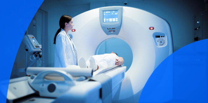
CT scan showing colon for screening colorectal cancer.
Computed tomography (CT) scans use low-dose radiation X-rays to capture internal images of the body. A CT scan creates multiple cross-sectional views, which are combined to form a highly detailed image for analysis. This is used to determine whether colorectal cancer has spread to other areas of the body, such as nearby organs or lymph nodes. CT scans may also be used to guide a physician if a biopsy of a mass is needed for evaluation.
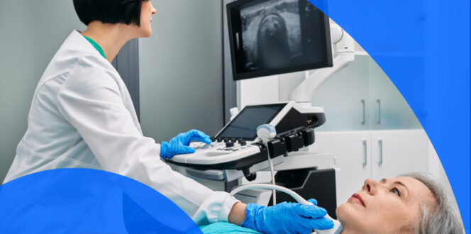
Ultrasounds use high-frequency sound waves produced by a handheld device called a transducer. The wand-like transducer creates these sonic waves, which the technician moves around the area being scanned. The waves bounce off internal structures and organs, creating images on a screen that are saved and then analyzed for possible diagnosis. For colorectal cancer, two types of ultrasounds are commonly used:
Positron emission tomography (PET) scans use a mildly radioactive tracer that is injected into the patient’s bloodstream. A gamma camera is then used to track this radiotracer and evaluate how this substance is taken up by tissues and bones, which can indicate whether cancer has spread or the efficacy of treatments. These highly detailed images offer a great deal of insight into cancer stages and behavior.
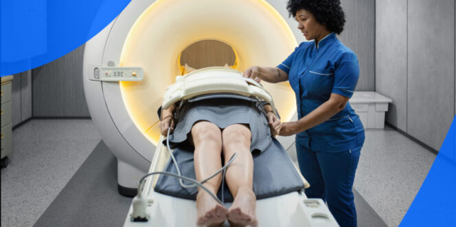
Magnetic resonance imaging (MRI) uses magnets and radio waves to create and capture internal images. For many patients, a contrast agent may be injected into the bloodstream to improve imaging detail, though this is not always necessary. Patients remain still in a tube-like MRI machine for the duration of the scan, which helps determine whether colorectal cancer cells have spread to other organs or structures outside the large intestine. Often, a thin wire-like device called an endorectal coil is inserted into the rectum during the scan to capture more detailed images.
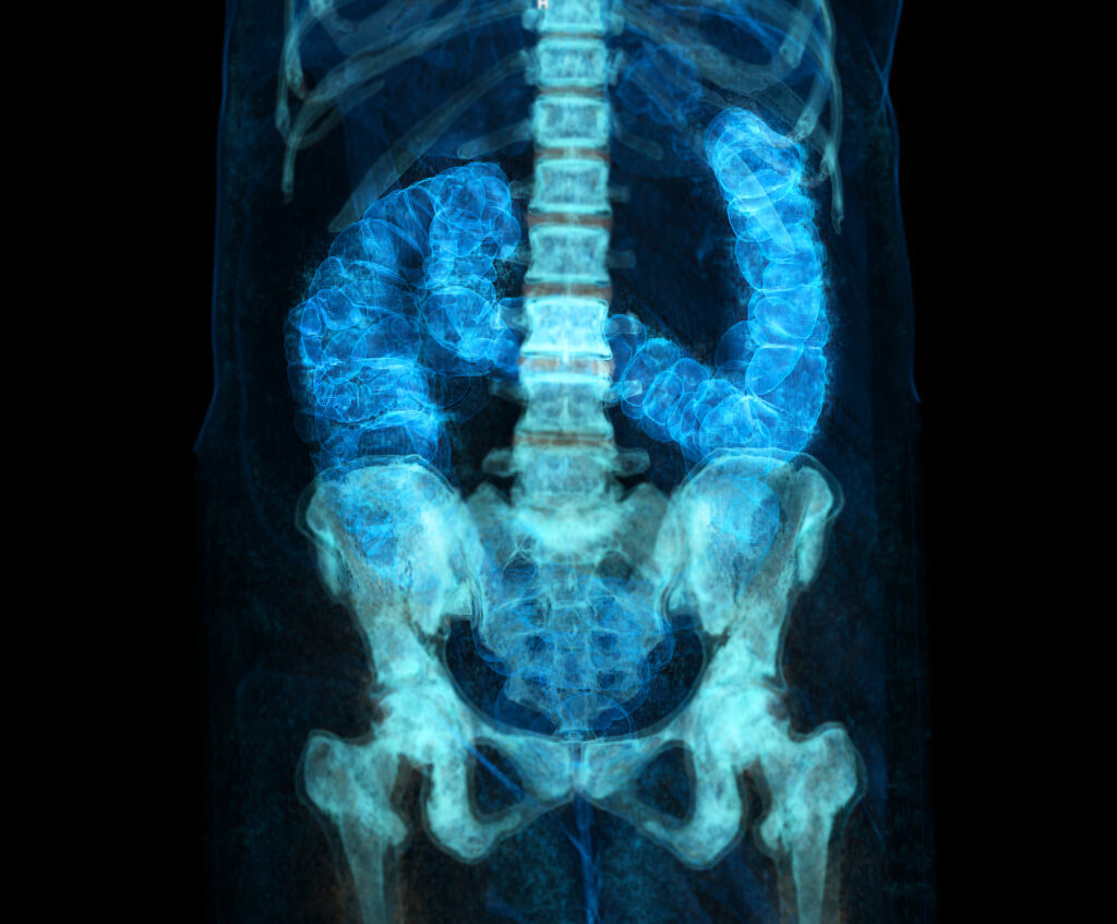
X-rays use low-dose radiation to create and capture internal images. They are more commonly used to determine whether colorectal cancer has spread to other organs or regions, rather than for identifying polyps or growths in the large intestine itself.
Cancer staging is a method used to determine how much cancer is in a patient’s body and how far it has spread. The staging classification helps guide treatment options and decisions about next steps. Here is a basic overview of what each stage means:
There are various tools and treatments for colorectal cancer, many of which depend on the stage of the cancer and other factors. Some common treatments include:
Turnaround times for colorectal cancer imaging results can vary depending on the facility and your doctor’s availability. Often, patients wait a week or more and receive their results during a follow-up appointment. With PocketHealth, you don’t have to wait as long—your results are available securely as soon as they’re uploaded, allowing you to review them before your follow-up visit.
When you do gain access to your results, it’s common to find medical terminology somewhat confusing, but Report Reader helps you to better understand your report by offering clear, straightforward definitions for medical terms—simply tap or click on any underlined words to reveal their meaning.
Here are some commonly asked questions regarding colorectal cancer.
Certain factors may increase a patient’s risk of developing colorectal cancer, such as having a family history of the disease or conditions like inflammatory bowel disease. However, the CDC suggests several lifestyle habits to help lower this risk, including:
This is a method to evaluate the large intestine for cancer or polyps, but it doesn’t require the insertion of an endoscope. Instead, a short tube is inserted about two inches into the rectum. Air is gently pumped into the large intestine through the tube to improve visibility. Like a traditional colonoscopy, bowel preparation is done in advance to eliminate obstructions and allow for better visibility.
A CT scan is then used to capture multiple images of the large intestine, which are evaluated for polyps, growths or other abnormalities. The pros of a virtual colonoscopy are that it is safer for patients with bleeding disorders or issues with anesthesia. The cons are that it may miss very small polyps. Additionally, if a polyp is detected, a separate colonoscopy will be needed to remove it, as a virtual scan cannot facilitate this.
The medical equipment used during a colonoscopy can also remove small polyps during the procedure, with other techniques employed for larger growths. The polyp(s) are removed and sent to a pathologist for analysis. If they show precancerous or cancerous traits, next steps may include watchful waiting, additional testing or initiating treatments. It’s important to remember that only 5 to 10% of polyps become cancerous. The majority are benign or can be removed before they turn cancerous. Often, when patients have polyps, more frequent screening colonoscopies are performed to monitor any changes over time.
A stool test is a diagnostic starting point, but it doesn’t definitively confirm cancer (or rule it out). The presence of blood in a stool test may suggest that a colonoscopy should be performed to look for possible polyps or other causes. However, there are many benign reasons for blood in a stool test, such as hemorrhoids, ulcers or other noncancerous conditions.
For anyone facing a possible colorectal cancer diagnosis, the treatment process and next steps can feel overwhelming. Seeking support during this time can be especially helpful. Your doctor will likely have a list of local support groups and resources. Additionally, here is a list of online support resources, including guides on how to navigate transportation, finances and lifestyle changes related to cancer treatment:
PocketHealth makes it simple to keep track of your scans and other medical results. All of your imaging records are in one secure location and can be accessed online anytime. If needed, reports can also be easily shared with other physicians in your care team. When used in conjunction with your medical provider’s professional advice, it is a powerful tool for organizing and understanding your imaging results and your health.
Another tool from PocketHealth is MyCare Navigator, which provides personalized insights to your health and identifies any recommended follow-up steps. This feature can also generate individualized questions to ask your doctor based on the findings in your report, ensuring you make the most of your consultation.
Published: February 28, 2025
Trusted by more than 900+ hospitals and clinics.