Lung cancer is one of the most common types of cancer worldwide. While there are various potential causes, smoking is the leading factor, responsible for about 85% of all lung cancer cases. Secondhand smoke is also a contributor, increasing the risk of developing the disease by 20 to 30%.
This article provides a brief overview of the condition, focusing specifically on diagnostic methods like medical imaging, biopsies and molecular testing. Screenings for high-risk patients and early detection can significantly improve outcomes, making diagnostic techniques a crucial part of cancer care.
Lung cancer develops when abnormal cells grow and spread out of control within the lungs, sometimes spreading to other areas of the body, such as other organs or bones. These uncontrolled cells may form tumors that can impair organ function and significantly impact health.
Cancers that originate in other parts of the body can spread to the lungs, but they are still classified based on their original site, such as metastatic breast cancer. For a diagnosis of lung cancer, the disease must begin in the lungs, either in the airways (the bronchi or bronchioles) or in the alveoli, the small air sacs within the lungs.
There are multiple types of lung cancer, which play an important part in diagnosis and treatment:
Despite its strong link to patients who smoke, lung cancer can also affect non-smokers. Here are the most common causes for this disease:
Early cases may not display symptoms, at first, but some common lung cancer symptoms patients may experience include:
These symptoms can have many possible causes other than lung cancer. If you have concerns, consider speaking with your doctor to schedule a personal health assessment or seek medical advice.
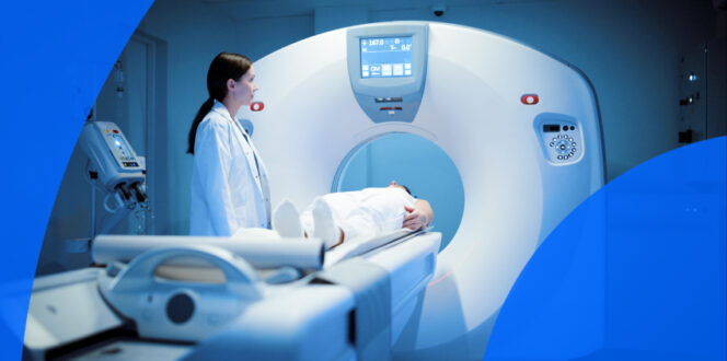
A CT scan of the chest
There are multiple methods to help physicians diagnose lung cancer. Here is a brief overview of each.
The initial physical exam is typically the starting point, with additional diagnostic methods used alongside it. During this appointment, the physician will likely ask about smoking habits, exposure to pollutants, family health history and any symptoms.
They will likely also listen to the lungs with a stethoscope to assess breathing, check the collarbone and neck area for swollen lymph nodes, take blood pressure and pulse readings and possibly compare the patient’s current weight to previous recordings. The doctor may also collect a mucus sample called sputum cytology, which involves the patient coughing up mucus from the lungs. It can then be tested for signs of cancer cells in a laboratory.
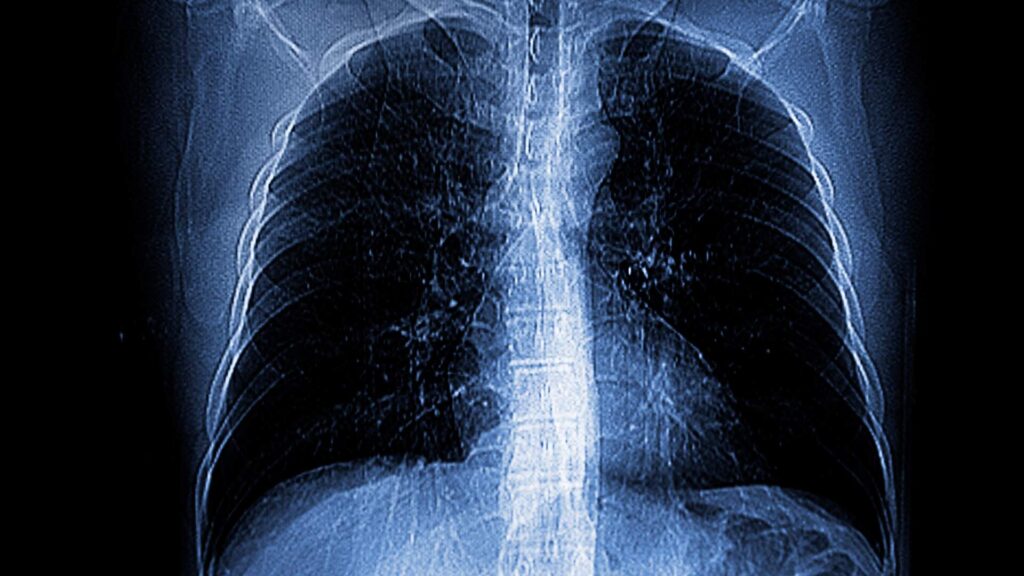
One of the first imaging tests commonly used is a chest X-ray. X-rays use low-dose radiation to capture images of internal structures, such as organs and bones. They may reveal tumors, changes in the lungs or spots suspicious for cancer. However, X-rays aren’t as detailed as other imaging methods. They are often a starting point for diagnosis, with more detailed imaging techniques used if there are signs of lung cancer in the initial X-rays.
A computed tomography (CT) scan also uses X-ray technology to create internal images. However, unlike a traditional X-ray that captures only a couple of images, a CT scan takes several and combines them into a highly detailed “slice” of the area being assessed, such as the lungs.
Because of this level of detail, CT scans can detect lung tumors that X-rays might miss, especially if they are small or in the early stages. It can also show if cancer has spread to nearby lymph nodes or other parts of the body.
Positron emission tomography (PET) scans are often performed alongside a CT scan, especially if lung cancer is suspected. For this imaging technique, a radiotracer called fluorodeoxyglucose (FDG) is injected into the patient’s bloodstream. This low-dose radioactive sugar tends to accumulate more in cancer cells, which absorb larger quantities of the radiotracer.
A gamma camera is used to detect areas of the body that collect more FDG, helping to determine if cancer is present and if it has spread to other regions. Both CT scans and PET scans are valuable for diagnosing and staging lung cancer, which will be discussed in more detail later in this article.
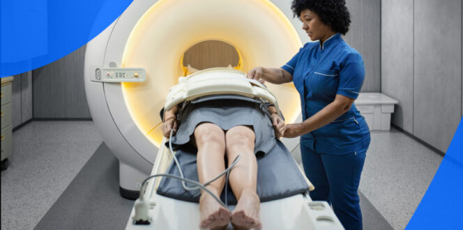
Magnetic resonance imaging (MRI) scans can also diagnose lung cancer. MRIs use radio waves and magnets to create highly detailed internal images of the body, including the organs, bones and tissues. They can help detect signs of cancer and determine if it has spread to other areas of the body.
One of the most definitive ways to diagnose lung cancer is through a biopsy, which is a tissue sample taken from the lungs. There are multiple methods for obtaining this sample, including:
If a biopsy confirms a cancer diagnosis, the tissue sample may also be tested for gene mutations and specific biomarkers. Different findings may require different treatment methods, depending on the specific mutation, proteins and other factors. Targeted therapy involves selecting medications or treatment types based on these biomarkers to ensure the most effective drugs are used to fight cancer cells.
Staging plays a crucial role in lung cancer diagnosis. It helps determine whether cancer cells have spread to other areas of the body, such as lymph nodes and organs, and can guide treatment decisions. MRIs, CT scans, and PET scans are all methods that can help determine this staging. The stages of lung cancer include:
Turnaround times for imaging results can vary widely depending on the facility and your doctor’s availability. Often, patients wait a week or more and receive their results during a follow-up appointment. With PocketHealth, you don’t have to wait as long—your results are securely accessible as soon as the report is available, allowing you to review them often before your follow-up visit.
Your imaging results are interpreted by a radiologist—an expert in medical imaging—who carefully reviews your scans and provides a detailed report of any findings. This report is then sent to your referring doctor, who, with a deeper understanding of your medical history, can offer further insights and recommendations.
To better understand your imaging reports, Report Reader provides clear, straightforward definitions for medical terms. Simply tap or click on any underlined words to reveal their meaning. This is paired with illustrations and highlights of anatomy in your imaging to help you better understand your results and prepare for follow-up appointments.
Here are some common questions regarding lung cancer that many patients may have.
It is not uncommon for early-stage lung cancer to have no obvious symptoms. However, as it progresses, many patients may experience a chronic cough, recurring pneumonia, hoarseness in the throat, difficulty breathing, chest pain and unintended weight loss. It is important to remember that these can also be signs of less serious conditions, and patients should consult with their physician if they have concerns. Because this disease can develop over years, high-risk patients may also want to ask their doctor about lung cancer screening options.
Aside from skin cancer, lung cancer is the second-most prevalent cancer type across all sexes. In those assigned female at birth, breast cancer is more common, while prostate cancer is more common in those assigned male at birth.
This disease tends to affect patients aged 65 or older more frequently than younger patients, and it is currently the leading cause of cancer deaths in the United States. However, these rates, along with new cancer case rates, are starting to decrease due to fewer people smoking and improvements in diagnostic and testing methods.
Vaping and e-cigarettes are still relatively new, so long-term studies on their chronic health effects have not yet been completed. While they typically don’t contain tobacco, they do have nicotine and other harmful chemicals. Some of these chemicals are thought to be carcinogenic, meaning there is a potential for cancer in the future, though more research is needed. What is known is that vaping can be harmful to the lungs, heart and other areas of the body.
Smoking is still, by far, the leading cause of lung cancer cases. However, in the United States, 10 to 20% of lung cancer cases occur in non-smokers. It is believed that many of these instances are due to second-hand smoke or exposure to pollutants like radon.
Regardless of the stage, receiving a lung cancer diagnosis can be overwhelming. Fortunately, there are numerous support resources available to help patients throughout treatment and beyond. Most doctors and specialists will have local resources to recommend, but many online supports may be helpful, including:
PocketHealth makes it simple to keep track of your lung imaging. All of your vital imaging is in one secure location and can be accessed online anytime. Reports can also be easily shared with other physicians in your care team, if needed. Additionally, it makes it easy to track health changes over time, such as with repeat imaging. When used in conjunction with your medical provider’s professional advice, it is a powerful tool for organizing and understanding your imaging results and your health.
PocketHealth MyCare Navigator gives personalized insights into your health and identifies any recommended follow-up steps. This feature can also generate individualized questions to ask your doctor based on the findings in your report, ensuring you make the most of your consultation. A lung cancer diagnosis may feel overwhelming, but working closely with your providers can give you the best chance of managing your condition and protecting your lung health.
Published: April 24, 2025
Trusted by more than 900+ hospitals and clinics.