If you have pain, numbness or tingling in your back, limbs or neck, you might be referred for a spine MRI. This article will explore what a spine MRI shows, how it is performed, how to prepare for your MRI scan, and what the results might look like.
After a spine MRI, you’ll likely want to know your results as soon as possible. PocketHealth gives you rapid, secure access to your medical images and results, often as soon as they’re released by the radiologist.
Magnetic resonance imaging (MRI) of the spine is an imaging test that captures images of the internal structure and tissues of your spine using a strong magnetic field and radio waves. Sometimes used in conjunction with other imaging tests like PET or CT scans, MRIs are particularly useful when examining soft tissues like the spinal cord or discs.
Your spine consists of bony vertebrae and soft discs and is divided into separate sections.
A spine MRI provides detailed images of the inner structures and workings of the different parts of your spine. Doctors use spine MRIs to diagnose, locate and monitor the treatment of several conditions, including:
When you check into the clinic, you’ll be led to a room where you can remove your clothes and jewelry and change into a gown. If you request a fast-acting sedative, this is when you’ll receive it. If you are having a contrast MRI, it will be administered, generally by IV.
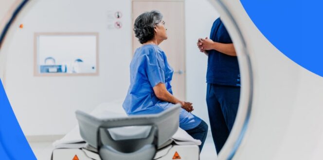
The MRI machine is a large cylindrical tube with an exam table that slides in and out. You must stay very still during your scan. The MRI technologist will position you on the table for optimal clarity and may use a device (called a coil) to keep you in place. Your exam table will slide into the circular opening. You’ll be given earphones to wear. The MRI scanner makes a lot of loud sounds as it works, banging and clanking as it scans, and the earphones should block out much of the noise.
You may be asked to hold your breath at points during the scan, or to shift position slightly. If your MRI involves contrast, you might feel warmth in the area being scanned. The MRI technologist will likely be in another room but will be able to hear and communicate with you through your headphones.
Your MRI scan can take anywhere from 30-120 minutes. If you had a sedative, you’ll need someone to drive you home afterwards.
If you’ve never had an MRI scan before, it might seem daunting. Knowing what to expect from the procedure can reduce any anxiety.
A spine MRI does not involve much preparation, but here are a few ideas to help you feel in control of the process.
Following these steps the day of your appointment will ensure your MRI scan is as seamless as possible.
The staff at the imaging clinic is not legally allowed to share the results of your scan with you. Below we’ll discuss who produces your results and how long it takes to receive them, and how to understand your MRI report.
The MRI technologist forwards your scan to radiologist for interpretation. The radiologist will send the images and a written report to your referring physician, who will share them with you.
Your doctor will discuss your MRI results with you at a follow-up appointment. The whole process can take a week or more.
With PocketHealth, you can access your results more quickly, from any device. You can also use MyCare Navigator to highlight any recommendations and create a list of personalized questions to help you prepare for your follow-up appointment.
You’ll get an official explanation of the findings in your MRI report from your doctor, but this section will explore what general scan findings look like.
With Report Reader, you can get simple, straightforward definitions of the complex medical terms in your report, so you’ll always be informed.
MRI reports contain a lot of medical terms and abbreviations. While we have a page to help you understand MRI terminology, this section will look at some of the more spine-specific clinical findings.
Your results will be generally classified as ‘normal/unremarkable’ or ‘abnormal’. Normal/unremarkable means just that: everything presented as usual, and no problems were detected. Abnormal results might indicate:
Here are some of the questions patients regularly ask about spine MRIs.
If required, brain and spine MRIs can usually be performed at the same appointment, though they will generally happen sequentially, rather than at the same time.
Your head is usually inside the machine during a spinal MRI.
MRIs are safe, non-invasive scans. Unlike X-rays, MRIs do not use radiation. If your MRI requires contrast, there is a very small risk of an allergic reaction to the contrast material. However, if you have kidney issues or are pregnant, discuss the risks of a contrast MRI with your doctor.
Spine MRIs are generally very accurate. MRIs are very effective at capturing images of soft tissue and distinguishing between areas of normal and abnormal soft tissue.
PocketHealth provides you with fast, easy access to your MRI results so you can see, store and even share your images with specialists in diagnostic quality. Having all your medical imaging in one place gives you peace of mind, and the confidence to advocate for your spinal health.
Published: February 6, 2025
Trusted by more than 900+ hospitals and clinics.