How does an MRI work? How long does an MRI take? Dr. Ania Kielar explains the ins-and-outs of this powerful technology.
Magnetic resonance imaging (better known as an MRI scan) is a medical imaging procedure that uses a strong magnetic field and radio waves to create detailed images of the body’s organs and soft tissues.
Dr. Ania Kielar, President of the Canadian Association of Radiologists and Vice-Chair of Radiology at the University of Toronto, explains how MRIs work, when and why they’re used, how long the procedure takes, when you can expect your results and how to access those results sooner with PocketHealth.
What’s an MRI and how does it work?
“Basically, you’re sitting inside a big magnet,” Dr. Kielar explains. “It very temporarily changes how your atoms orient inside your body, but it doesn’t change your DNA; it doesn’t change anything in your body.”
The magnetic field uses strong magnets to push the body’s protons out of regular alignment to align in the same direction. The MRI machine then receives radio waves that send those protons spinning back to their regular alignment.
Once the radio waves are turned off, a computer analyzes the energy released and the time it takes the protons to realign with the field to create detailed images of the area of interest. This amount of time is called T1 or T2 relaxation time.
MRI images are cross-sectional and can be taken in 3 different ways to capture a complete image of the body:
- Coronal. A cross-sectional front view that presents a mirror image of the body. For example, in a coronal view of an MRI scan, the left side of the body will be on the right side of the image, and vice versa.
- Sagittal. A cross-sectional view taken from a side-profile view of the body.
- Axial. A cross-sectional view that will also present as a mirror image. In an axial view of an MRI scan, images are taken from feet to head or bottom to top.
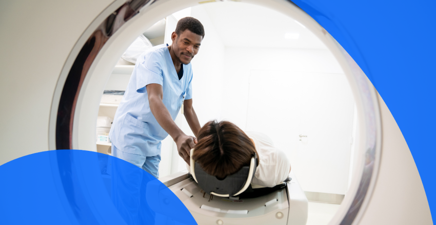
During an MRI exam, patients are required to lie face up on a bed that is threaded into a doughnut-shaped MRI machine similar in appearance to a CT scanner but longer and able to accommodate a larger portion of the body.
What’s the difference between an MRI and a CT scan?
MRIs and CT scans use different technology to provide clear images of the body’s inner structures. The reports generated by these machines help doctors diagnose conditions or monitor treatment.
An MRI exam creates a magnetic field in the body and captures images by assessing the movement of protons in that magnetic field, while a CT scan compiles multiple X-rays into a single, highly detailed image. MRI scans provide clear images of soft tissues like muscles, ligaments and tendons and can show blood vessels and blood flow to and from organs. CT scans are better at capturing defined images of the hard structures of the skeletal system, similar to an X-ray.
Your doctor will choose the imaging procedure best for your circumstances and will use the resulting detailed pictures and report to inform your treatment plan.
How long does an MRI scan take from start to finish?
MRI exams take anywhere between 30 and 60 minutes, and an MRI technologist will be close by at every stage of the procedure. MRI machines are not silent, and patients should be prepared to hear a lot of loud knocking and clicking during the MRI.
“Each sequence has its own sound, so you may feel like you’re in a dance club at times,” Dr. Kielar says. “They do put stuff over your ears, and they talk to you through earphones so if you’re uncomfortable you just have to tell the tech. We give you a button you can press, and we’ll pull you out immediately if you’re having any issues.”
What does an MRI show?
MRI images capture the details of different organs and soft-tissue structures, including:
- The brain and spinal cord
- The inner ear
- The heart and lungs
- Pelvic organs, like the bladder or uterus
- Blood vessels and the venous system
- Hip, ankle or knee joint tissues
- Muscles, ligaments and tendons
MRI scans can also indicate different conditions, like
- Multiple sclerosis (MS)
- Soft-tissue cancers, like breast cancer
- Spinal cord conditions or spine injuries
- Muscle, ligament or tendon damage
- Bone infections
- Heart conditions
- Vein obstructions and blood flow conditions
- Endometriosis
“The images we get are exquisite,” Dr. Kielar says. “They’re very useful for looking at the brain and very useful for looking at pelvic structures, such as the reproductive organs. They’re also very good for looking at soft tissue in general for, say, tumors in the bones, and they’re excellent for looking at the bones themselves.”
What are the different types of MRI scans?
Various types of MRI scans explore different areas of the body, including
Each detailed MRI scan provides valuable information about the organs and tissues in the region, including blood flow, inflammation or tissue damage.
MRI exams typically require a contrast agent, usually gadolinium, to change the magnetic properties of water molecules inside the body and improve image quality. The contrast material allows an MRI scan to capture detailed structures by creating higher contrast areas that are easier to assess.
“This is excreted through the kidneys,” Dr. Kielar says. “So, we need your kidneys to work reasonably, in the same fashion as the CT scanner needs your kidneys to work OK for the contrast agent. They’re different agents, though, so if you’re allergic to one, it doesn’t mean you’re allergic to the other. The chance of being allergic to gadolinium is very low.”
How can patients prepare for an MRI?
Your MRI scan gives your medical team information to help diagnose or assess your condition or treatment. An MRI scan is a simple, painless procedure, but some preparation is required before your appointment.
- Bring your requisition. Your requisition contains essential information for the imaging clinic, including a description of the area to be scanned and notes from your referring practitioner. Having the form on hand will make the check-in process easier.
- Wear comfortable clothing. You’ll be taken to a separate room to change into a hospital gown for your scan, so wear comfortable clothing you can easily change out of.
- Remove metal jewelry and body piercings. Metal items can be pulled toward the strong magnets used in an MRI. “When you’re making your appointment, they’ll ask your doctor to fill out a form to make sure you don’t have anything that’s dangerous inside a magnet,” Dr. Kielar says.
- Disclose any implants. The MRI may impact some metal implants. You should alert your doctor if you have an implant or worked with metal in the past, or if there’s a possibility any metal shrapnel or bullets are in your body. Your doctor will clarify whether you have any metallic implants prior to your MRI appointment, including some types of metallic joint prostheses, fillings and hearing aids, contraceptive devices or drug fusion pumps. “If you have a pacemaker or if you have cochlear implants or if you had brain surgery with clips, the technologist will ask you about that even before you arrive,” adds Dr. Kielar.
- Refrain from eating (in some cases). Some MRI scans require an empty stomach, but not all. Check your requisition form or confirm with the imaging clinic or hospital before your appointment if you’re unsure. Receiving contrast fluid may also mean you need to have an empty stomach. “If we’re giving you intravenous contrast, we don’t want the stomach to be full because some people feel unwell after getting the contrast agent,” says Dr. Kielar. “Not most, but sometimes.”
- Drink only clear fluids. Even if you’re not required to fast before your appointment, you should drink only clear liquids. And if your MRI requires a contrast agent, avoid caffeine for 24 hours before your appointment.
- Complete any necessary bloodwork. Bloodwork may be required if a contrast agent will be used for your MRI scan. The blood test ensures your kidneys are working properly and will be able to clear the contrast agent from your system.
- Clarify medication safety. While regular medications can generally be taken, Dr. Kielar encourages people to come prepared anytime they enter a healthcare facility. “Whenever you go to the hospital, I strongly recommend that you have a list of your past medical history and your medications.
Are there any other concerns or risks that accompany an MRI?
MRI scans are generally safe and don’t release radiation or damage surrounding tissue, but a small number of patients may experience minor discomfort during their MRI scan, due to:
- Anxiety or claustrophobia. Patients who have a fear of enclosed spaces should share any concerns with their doctor or radiologist before their MRI scan. Your doctor can likely improve your MRI experience if you have anxiety. “There is one to two percent of the population that will be claustrophobic and feel a little bit uncomfortable in there, so they can get Ativan or other medications to kind of make them a little bit calm or sleepy,” Dr. Kielar says. “If you do have to take those medications then you cannot drive so you will need someone to drive you home.”
- Muscle twitching. Most MRI machines operate at a strength of 1.5 Tesla, but some operate at double the strength, or 3 Tesla. “Sometimes, this can cause your muscles to twitch a tiny bit,” Dr. Kielar says. “It’s normal but good to know in advance.”
- Tattoos. Body art can be a problem because some tattoo ink contains magnetic particles that may heat up inside the powerful magnetic field used for the scan. “We’ll warn you that if you do start feeling hot on your skin where your tattoos are, you have to tell us, and we’ll take you out immediately.”
- Pregnancy concerns. It’s crucial to inform the radiologist if you are pregnant or suspect you might be. “We try not to scan people who are pregnant,” says Dr. Kielar. “If we can avoid it until later, we will.” However, an MRI can be performed on a pregnant patient under specific circumstances. “If we need to do it, we will do it,” says Dr. Kielar, but generally doctors will avoid any sort of imaging on pregnant women until after the baby is born. Dr. Kielar also advises that intravenous contrast dye not be given during pregnancy.
- Allergy. According to Dr. Kielar, some patients may feel flushed or warm after being given contrast fluid but fewer than one percent of patients will have an allergic reaction requiring medical treatment. If you’re concerned, discuss any allergies with your doctor before your MRI scan.
What should patients do after an MRI?
MRI scans are non-invasive and generally do not require recovery time. Patients can resume regular activities after the scan, including taking pain medication.
“If we use a contrast agent, we usually advise patients drink a reasonable amount of fluid after the exam,” Dr. Kielar says. Drinking 6–8 glasses of water after your scan will help clear the contrast dye from your system.
How to understand your MRI results
Your MRI scan results will indicate areas of concern by showing the radiologist different cross-sectional images of your body and combining those images or “slices” into one complete view.
Your healthcare provider will send the radiologist any of your medical history that will help in assessing your MRI images. The radiologist will then examine the images and create an MRI report detailing what they see.
Can MRI results be seen immediately?
MRI results are usually available within a few days if you visit an imaging clinic but can be ready sooner during a hospital stay or within hours in an emergency.
Your MRI results may be available online through your healthcare provider’s patient portal or the imaging clinic, but you can access your MRI results quickly with PocketHealth.
PocketHealth securely stores all your medical imaging and reports, so you can access and review your results as quickly as possible. Access your records today.
Understanding common terms in your MRI report
The radiologist may use terms that are unfamiliar to you, but this list can help.
- T1 weighted. A type of MRI image that uses the T1 relaxation time of the tissues to create images. T-1 weighted images allow the radiologist to view soft tissues and can help identify tumors or other lesions.
- T2 weighted. A type of MRI image that uses the T2 relaxation time of the tissues to create images. T-2 weighted images allow the radiologist to view fluid-filled space and can help identify inflammation, swelling or fluid buildup.
- Signal intensity. Indicates the darkness or brightness of an area on an MRI image. High-intensity areas appear white, medium-intensity areas appear grey and low-intensity areas appear black. Signal intensity can show the difference between normal and abnormal tissue, helping the radiologist identify areas of concern.
- Enhancement. An increase in signal intensity in a specific area following contrast fluid injection. Enhancement can show changes to tissue, including increased blood flow.
- Lesion. An area of abnormal tissue that appears different from surrounding tissue. Lesions can be caused by different factors, including infection, inflammation or a serious illness like cancer.
- Edema. Inflammation or swelling caused by an accumulation of fluid. Edema shows up as an area of increased signal intensity on an MRI and can indicate injury or infection.
You can also learn more about the medical terminology used in your MRI report by using PocketHealth Report Reader and reviewing this article.
Become a confident advocate for your health
Understanding your MRI report allows you to confidently discuss your healthcare decisions and become an informed participant in your care.
Knowing how to prepare for your MRI appointment and what to expect, what to do after your scan and how to understand your MRI scan results lets you take more control of your healthcare journey.
PocketHealth is here to help you access your MRI results quickly. Having secure access to your images and reports lets you understand those results ahead of an appointment with your referring physician so you can prepare the right questions and feel confident at your appointment.
How PocketHealth works
Learn more about how to use PocketHealth to access and share your MRI results.
Published: February 1, 2022