Preparing for an MRI: What to Wear, What to Avoid and Other Tips
July 25, 2024
Read More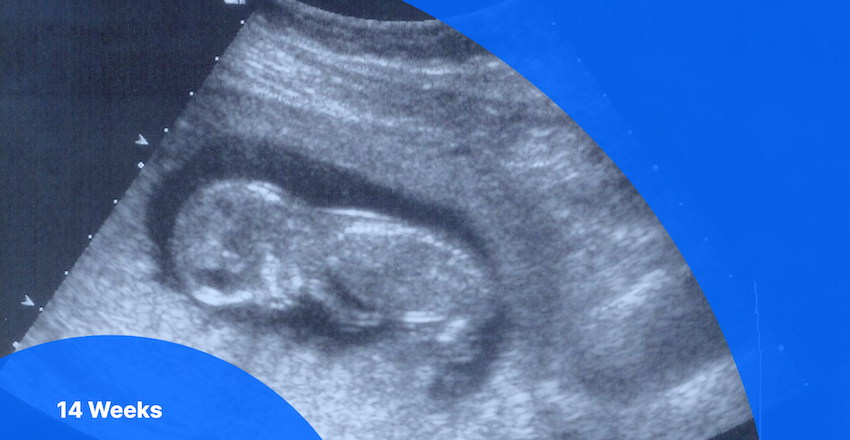
By 14 weeks pregnant, you’re officially in your second trimester! Your baby is now fully formed and has even started to develop hair (also known as lanugo) to keep warm as it continues to grow in the coming months.
If you did not have prenatal ultrasounds between weeks 11 and 13, your practitioner will send you for one in week 14 to screen for any chromosomal anomalies. Miscarriage rates also drop significantly in the second trimester compared to early pregnancy, according to the University of California Department of Obstetrics and Gynecology.
If you getting ready for your 14 week scan, read on to find out:
Your practitioner won’t likely send you for a 14 weeks pregnant ultrasound unless you didn’t have a scan between 11 and 13 weeks. During this scan, the sonographer or technician will be screening for:
If you didn’t have a scan between 11 and 13 weeks, your ultrasound technician may also screen to determine:
A nuchal translucency (NT) screening is not a diagnostic test, but a measurement of the thickness of the area of fluid buildup at the back of the baby’s neck. Some fluid is expected, but too much might indicate a potential risk of chromosomal anomalies like Trisomy 21 (i.e., Down syndrome).
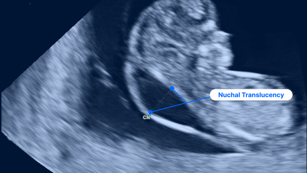
Pregnancy ultrasound image showing increased nuchal translucency
If your NT test comes back inconclusive or positive, you have the option to request more information about the results. You can also ask your practitioner to discuss further testing with you, including:
Diagnostic testing is also available and includes the following options:
You can ask your practitioner what information these tests can give you and whether they recommend one test over another. You can also choose not to receive more information at this time and wait until your next ultrasound between 18 and 20 weeks.
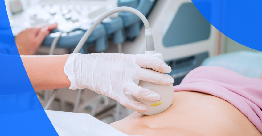
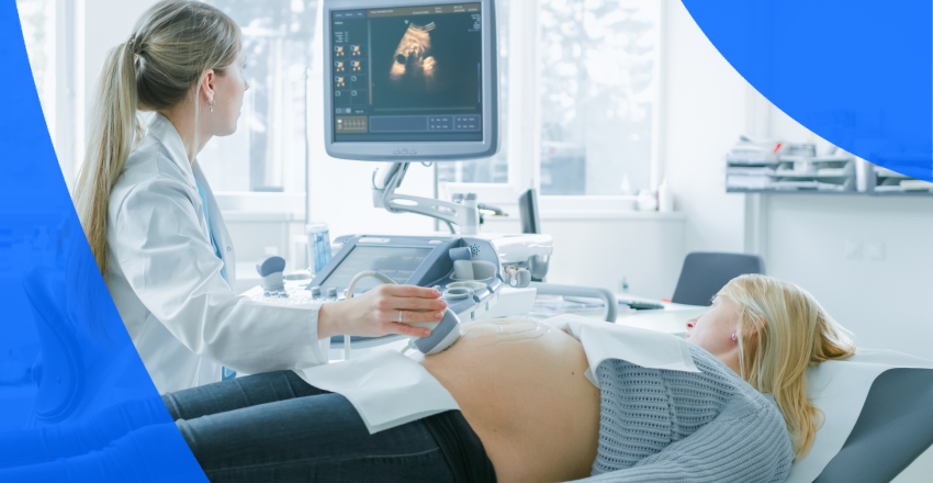
During a medical ultrasound, a sonographer or trained technician uses a hand-held device called a transducer to make sound waves (too quiet for human ears to hear) that travel painlessly through your body to your baby. An ultrasound machine can detect the sound waves and use them to create an image of your baby’s exact size, shape and position.
Unlike X-rays, ultrasounds don’t use radiation to capture images and are safe when performed by a trained sonographer or ultrasound technician, according to the U.S. Food & Drug Administration. Only practitioners trained to give fetal ultrasounds can give you a pregnancy ultrasound.
You’ll most likely receive a transabdominal ultrasound at your 14 weeks appointment. During a transabdominal ultrasound, you’ll be asked to change into a gown and lie down on your back on a reclined, cushioned table. The sonographer will place gel on your belly and abdomen and slide a transducer across the gel to capture detailed images of different parts of your womb, cervix and the developing fetus.
Pregnancy ultrasounds require some preparation. You can make sure you’re ready for your 14 weeks transabdominal ultrasound by:
Using the results of your 14 weeks ultrasound, your healthcare practitioner will learn valuable information about how the fetus is developing and how your body is changing. This week, the fetus will be about 3.4 inches (8.5 cm) from head to bottom.
By this stage of pregnancy, biparietal diameter (measures the developing skull of the fetus) will also be taken to better determine fetal weight and age. The baby’s head will begin to round out and become more proportionate to the length of its body as its body, as its arms and legs continue to grow. Ovaries or testes will be fully formed, and genitalia is now becoming distinct on the outside of the body. Your baby can also turn its head and even bring its fingers to its mouth.
Although you won’t be able to see it on your ultrasound, your baby will be developing muscles for swallowing and will be testing them out. The fetus will also begin to grow fine hair (lanugo) and develop unique fingerprints. You may notice that the yolk sac has disappeared—it’s been absorbed by the fetus for nourishment.
Though sometimes used as synonyms, sex and gender mean different things. Sex refers to physical characteristics and attributes, while gender refers to the social roles individuals use to identify themselves in the world. Predictions of sex are fairly accurate at 14 weeks. By now the baby’s genitalia have developed internally and have become much more distinct externally, but it is common to wait until around to determine the sex of the baby during your anatomy scan ultrasound between 18-20 weeks.
However, your doctor will likely be able to see the genital tubercle, or the very beginnings of genitalia forming. Called the nub theory, by 13 weeks, it’s over 98% accurate in predicting sex by examining the angle of the tubercle. And accuracy continues to increase with gestational age.
At your 14 weeks ultrasound appointment, the ultrasound technician can’t legally answer questions about what can be seen in your ultrasound or discuss the results of your ultrasound report. The technician will prepare images, which are reviewed by a radiologist. The radiologist then prepares a report for your practitioner to discuss with you at your ultrasound follow-up appointment.
Your healthcare practitioner will go over the results of your 14 weeks pregnant ultrasound with you at your follow-up appointment. The ultrasound provides insight into the health and development of the fetus and your own health, so make sure to ask any questions you may have, including this list to get you started.
You may be eager to access your 14 weeks pregnant ultrasound images and report as soon as possible. With PocketHealth you can quickly and easily access and share your pregnancy ultrasound images and report–often before your follow-up appointment with your practitioner. Access your records here.
PocketHealth enables you to securely access, share and store your imaging and other health information in one place. You can also easily share images with family and friends and see how your baby is changing over time.
Pregnancy ultrasound terminology can be complicated, but PocketHealth Report Reader is there to help. Report Reader makes it easier to understand terms in your ultrasound report and feel more prepared and confident when speaking to your pregnancy care practitioner.
Your 14 weeks pregnant ultrasound will track your baby’s development, using biparietal diameter measurements for the remainder of the pregnancy. If you didn’t have an ultrasound between 11 and 13 weeks, your practitioner will also measure nuchal translucency.
Being prepared for your 14 weeks pregnant ultrasound can help you feel comfortable during your appointment and confident when speaking with your practitioner. The more knowledge you have, the more empowered you’ll be as you continue through your second trimester.
Learn more about how to use PocketHealth to access and share your pregnancy ultrasound records.