This article was reviewed by Dense Breasts Canada for accuracy.
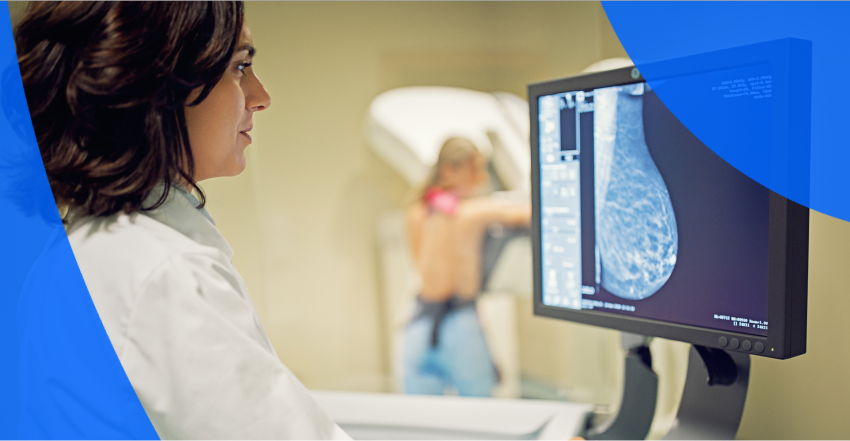
Mammograms are an excellent breast cancer screening tool because they can detect signs of cancer early, often before any lumps can be felt or other noticeable symptoms arise.
A mammogram is the only way breast density can be determined. Usually, the radiologist makes the decision by looking at the images, but there is also software that can now measure density. Depending on how dense your breast tissue is, you may benefit from additional imaging to find cancer not seen on the mammogram. Understanding your breast density is important, as dense breasts have an increased risk of breast cancer, and can also cause a masking effect, making signs of cancer harder to see on a mammogram. This article will explain:
Breast density describes the composition of your breast tissue. All breasts contain both fatty tissue and fibrous or glandular tissue (also called fibroglandular tissue). Dense breasts contain more fibrous and/or glandular tissue than fatty tissue.
Dense breasts are very common and perfectly normal. Younger women are more likely to have dense breast tissue, with 56% of women in their 40s having dense breasts, compared to only 27% of women in their 60s.
Having dense breasts can hide cancer on a mammogram. Fat looks black or dark gray on a mammogram. Fibrous and glandular tissue both look white in mammogram images—and so does cancer. In dense breasts, the increased fibroglandular tissue can have a potential masking effect, obscuring signs of cancer.
Breast density is not the same as firmness. You cannot assess breast density by look or feel. Radiologists examine mammogram images to determine breast density and use the Breast Imaging Reporting and Data System (BI-RADS) to classify the level of dense breast tissue, which is broken down into the following 4 categories:
For screening purposes, both categories C and D are considered dense breasts.
In the U.S., 38 states and the District of Columbia are required to provide some level of breast density notification after a mammogram. The FDA also recently announced a national requirement to notify patients and referring providers about breast density after mammogram screenings, that dense tissue can hide cancers and that supplemental screening may find many of those hidden cancers. However, this won’t be fully in effect until late 2024 and will only report a “dense” or “not dense” category.
In Canada, 7 provinces have already made reporting and notifying all patients about their breast density results a requirement. Reporting density results to all patients is not yet a requirement in Newfoundland & Labrador, Northwest Territories, Saskatchewan and Yukon, although women with over 75% density (Category D) are asked to return for annual mammograms. Newfoundland and Labrador and Saskatchewan are committed to providing density notification to all women in the future.
If you find out you have dense breasts, it’s best practice to check in with your primary care physician about appropriate mammogram screening frequency and whether supplemental imaging is available in your state, province or territory.
The two biggest risk factors for breast cancer are age and being female. Breast density also poses another risk. Having Category C or D dense breasts can be even more of a risk factor than your family health history. A study of more than 1,100 matched pairs of women found that women with extremely dense breasts (i.e., Category D) are 4-6 times more likely to develop cancer than women with Category A density. The denser the breast, the higher the risk.
In addition, it can be more difficult to detect cancer in dense breasts, since the fibroglandular tissue can mask or obscure signs of cancer. This means cancer may not be found until a more advanced stage, making the prognosis potentially more severe and treatment more intense.
Regular mammograms are an important tool for breast cancer screening. In the U.S., new draft guidelines proposed this year by the U.S. Preventive Services Task Force (USPSTF) recommend that all women have regular mammogram screenings every other year beginning at age 40. There weren’t any dense breast-specific changes made to the USPSTF draft guidelines.
In Canada, the starting age for screening without a referral depends on which province or territory you live in. For example, British Columbia, Nova Scotia, PEI and Yukon all begin breast cancer screening at age 40, when women can refer themselves, without a doctor’s requisition. Sometime in 2024, both Ontario and New Brunswick have committed to lowering the screening age to 40. The remaining provinces and territories (except for Nunavut, which doesn’t have a screening program) start at age 45 (only Alberta) or 50 but accept women over 40 with a requisition.
For Canadians with dense breasts, screening recommendations differ depending on the province. For example, under the Ontario Breast Cancer Screening Program, only women with Category D density are asked to return annually for a mammogram. However, women with Category C density (and any woman aged 50-74) can self-refer annually for screening.
For more details on your specific province or territory, Dense Breasts Canada has collated the provincial and territorial guidelines for comparison. Canadian mammogram screening guidelines also emphasize that the decision to have a mammogram is yours. If you have dense breasts, or any other concerns, and are under age 50, you can ask for a referral.
“Early breast cancer detection is key to saving lives, which is why it’s important to start mammogram screening at age 40. If you cannot self-refer in your region, you can request a requisition for a mammogram from your family doctor. Since dense breast tissue can mask cancer on a mammogram, supplemental screening, like breast ultrasounds and MRIs, also plays a critical role in cancer detection. If supplemental screening isn’t easily accessed in your region, ask and advocate for yourself.”
– Dense Breasts Canada
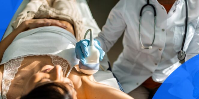
Mammograms are the most widely available breast cancer screening tool for all women in the U.S. and Canada. Depending on your location and density category, you may also be eligible for screening ultrasounds. For example, B.C. and Alberta offer supplemental screening ultrasounds for women with Category C or D breast density, but these require a requisition.
Additional imaging may be ordered to further investigate and diagnose any abnormal findings found on a mammogram. Here’s a breakdown of 4 types of diagnostic imaging available in the U.S. or Canada:
Your ability to access supplemental screening or diagnostic imaging will depend on your location, mammogram result and a requisition from your referring physician. It’s best to confirm with your healthcare provider what is available to you in your state or province. If you’re having any issues getting a requisition, try these tips from Dense Breasts Canada.
How, and whether, you receive your density results depends on your location.
U.S.
Canada
Turnaround times for imaging results can vary widely depending on the facility and your doctor’s availability. Often, patients wait a week or more and receive their results during a follow-up appointment. With PocketHealth, your imaging results, including your breast density category, are securely accessible as soon as they’re approved for release by the hospital or imaging clinic. This allows you the opportunity to review your results and prepare questions ahead of your follow-up visit.
If you’re part of a regular breast screening program, you’ll receive a letter in the mail after your mammogram containing your results and dense breast category (i.e., A, B, C or D). If your results are normal, your letter will say so and will let you know when to get your next screening. If you have a family doctor, they will also be notified.
Keep in mind that a normal mammogram means no cancer was seen. If you have category C or D dense breasts, your risk profile is higher, so it’s best to play it safe and ask for supplemental screening.
An abnormal result does not mean you have cancer, but it does indicate you will need further testing. Your doctor will be notified of the results and will set up a follow-up appointment. If you don’t have a primary care provider, you’ll be assigned one by the screening program. Since abnormal findings are included in the radiologist’s report, you can also access them through PocketHealth.
Mammogram images may identify several types of abnormal findings, including:
The findings in your screening mammogram will fall into 2 categories:
To help you better understand your breast imaging results, PocketHealth provides clear definitions and illustrations for complex medical terms—plus an in-depth explanation of your full imaging report. This is paired with highlights of key anatomy in your imaging to help you better comprehend what you’re looking at.
Doctors use BI-RADS to explain the findings in your diagnostic mammogram results. Results are reported under the following 7 categories:
If you have Category C or D dense breasts, it’s important to discuss supplementary imaging options with your healthcare provider. But you’ll also want to keep yourself informed and up-to-date. Here are some resources to help you get started:
American resources:
Canada resources:
Breast cancer screening mammograms save lives. They can detect cancer at the earliest stages, making treatment options easier to implement and drastically improving breast cancer outcomes overall.
If you’re located in Ontario, you have the option to get your breast cancer risk score and determine your eligibility for the Ontario Breast Screening Program (OBSP), as part of PocketHealth’s suite of breast health tools. If you’re eligible for OBSP, you can even book your next mammogram directly through PocketHealth.
PocketHealth makes it simple to keep track of your medical imaging results. All of your images and reports are permanently available in one secure location and can be accessed online—anytime, anywhere. Reports can also be easily shared with other members of your care team, if needed. When used in conjunction with your medical provider’s professional advice, it is a powerful tool to better understand your imaging results.
PocketHealth also provides personalized health insights based on the findings in your report to help you stay on top of any next steps. This includes clearly surfacing any follow-up actions found in your report and generating customized questions to ask your doctor so you can make the most of your follow-up appointment.
If you have dense breasts, you may require supplemental breast imaging, like a 3D mammogram or ultrasound, to take advantage of the life-saving potential of early detection. When you stay informed about your breast density you can be in control and proactive when it comes to maintaining your breast health.
Learn more about how to use PocketHealth to access and share your mammogram images, report and breast density results.
Published: November 27, 2023
Trusted by more than 900+ hospitals and clinics.