A breast ultrasound is an extra imaging exam doctors recommend in some cases to assess internal breast health. You might be referred for this non-invasive scan to provide more information after a mammogram or physical exam.
After you have a breast ultrasound, the clinic will send your images and report to your referring physician, who will discuss them with you. That follow-up process can take several days, sometimes even longer than a week. If you want faster access to your results, PocketHealth can help. With PocketHealth, you can get secure early access to your medical images and reports, often as soon as they’re released.
A breast ultrasound uses sound waves to create and capture live images of the inside of your breast. Safe and non-invasive, a breast ultrasound provides doctors with a different perspective and view of your internal breast tissues and structures.
Doctors use breast ultrasounds to gain more clarity and information about your breast health and to further examine any abnormalities found in a physical breast exam or screening mammogram.
You might be referred for a breast ultrasound:
A breast ultrasound uses high-frequency sound waves to capture images of your internal breast tissue, while a mammogram uses low-dose X-rays.
Mammograms are an effective method to detect cancer in the earliest stages, which improves both treatment and recovery. If a mammogram is unclear, or shows an abnormality, a breast ultrasound is often used to provide more information.
The two imaging tests are not competing, but complementary. A mammogram excels at finding anomalies and detecting signs of cancer early, which is critical to positive health outcomes, while a breast ultrasound provides insight into the nature of any abnormal findings.
A mammogram examines your entire breast and provides a record of any changes in breast tissue over time. However, a breast ultrasound can provide more details about a specific area of your breast and distinguish whether a lump is a fluid-filled cyst or a solid mass.
Being referred for a breast ultrasound scan after a mammogram is a fairly common occurrence. It does not automatically mean something is wrong. An ultrasound scan gives your health care team better visibility into any areas that were previously unclear.

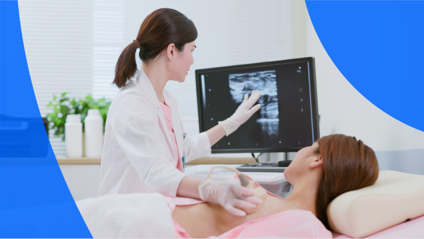
During your breast ultrasound, you’ll undress from the waist up and lie on an exam table in the scanning room. The sonographer will help arrange you into the best position, usually with your arms raised above your head. You may be asked to shift positions during the scan.
The ultrasound technician will apply a conductive gel to your skin, which helps transmit the high-frequency sound waves into your breast tissue. The technician then glides the transducer over the gel, holding it in different spots and angles to capture the best images. They will be examining a specific area, not your entire breast.
You will feel some pressure from the transducer, but the procedure should not be painful. A breast ultrasound usually takes about 30 minutes. Afterwards, you can wipe off the water-based gel and go about your day as normal.
Very little preparation is needed for a breast ultrasound. When you book your appointment, check with the imaging clinic for any special instructions.
What to do:
What not to do:
Many women report having sensitive breasts before the start of their menstrual cycle. If that describes you, the most opportune time to schedule a breast ultrasound is when your breasts are less sensitive and tender, usually the week after a menstrual period.
The ultrasound technician is not legally allowed to discuss the results of your scan with you. They will send your images to a radiologist—an expert in medical imaging—who carefully reviews your scans and provides a detailed report of any findings. This report is then sent to your referring physician.
Turnaround times for imaging results can vary widely depending on the facility and your doctor’s availability. Often, patients wait a week or more and receive their results during a follow-up appointment. With PocketHealth, your ultrasound imaging results are securely accessible as soon as they’re approved for release by the hospital or imaging clinic. This allows you the opportunity to review your results and prepare questions ahead of your follow-up visit.
The radiologist will interpret the results, and send them to your doctor who has a deeper understanding of your medical history and past exams. Your doctor will then offer further insights and recommendations on next steps in a follow-up appointment.
To make the most of your follow-up appointment, PocketHealth also provides personalized health insights based on the findings in your report and generates customized questions to ask your doctor such as:
A breast ultrasound uses the Breast Imaging Reporting and Data System (BI-RADS) to report your results. BI-RADS assigns categories to each finding, from 0-6. The lower the category number, the lower the probability the finding is cancerous.
To help you better understand your breast ultrasound results, PocketHealth provides clear definitions and illustrations for complex medical terms—plus an in-depth explanation of your full imaging report. This is paired with highlights of key anatomy in your imaging to help you better comprehend what you’re looking at. For more information, check out how to understand an ultrasound image and how to read your mammogram results, which includes a complete explanation of each BI-RADS score.
An abnormal breast ultrasound
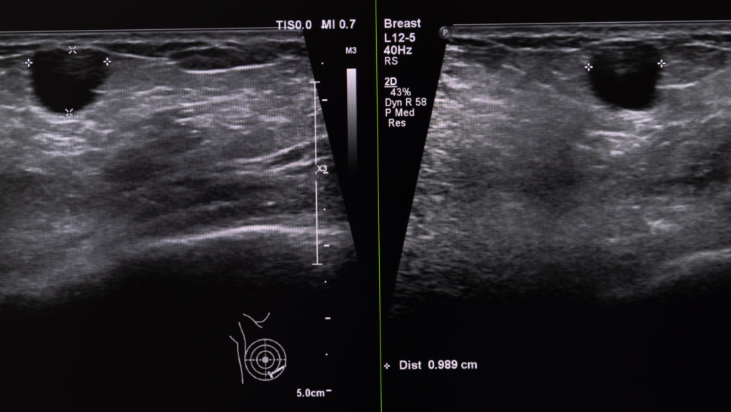
Abnormalities can have many different appearances, depending on the underlying cause. A lesion might be a calcification, a cyst, a mass or even an infection, each of which looks slightly different on an ultrasound.
A cyst is a fluid-filled sac. On a breast ultrasound it will look like a round black shape with thin walls.
An ultrasound is not a screening test for cancer, but it can distinguish between a fluid-filled cyst (unlikely to be cancer) and a solid mass (higher possibility of being cancerous). A breast ultrasound can eliminate or indicate the need for further testing.
A mass is solid, while a cyst is filled with air or liquid. Cysts appear darker on ultrasound images and have rounded shapes. Solid masses are often lighter in color, with an irregular outline.
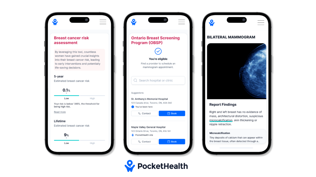
PocketHealth’s breast health tools provide patients with personalized breast cancer risk assessments and simplified mammogram management. Designed to increase early detection of breast cancer, these tools enable patients to take control of their breast health. Learn more about PocketHealth’s breast health tools here.
PocketHealth makes it simple to keep track of your medical imaging results. All of your images and reports are permanently available in one secure location and can be accessed online—anytime, anywhere. Reports can also be easily shared with other members of your care team, if needed. When used in conjunction with your medical provider’s professional advice, it is a powerful tool to better understand your imaging results.
PocketHealth also provides personalized health insights based on the findings in your report to help you stay on top of any next steps. This includes clearly surfacing any follow-up actions found in your report and generating customized questions to ask your doctor so you can make the most of your follow-up appointment.
Aileen used PocketHealth to stay on top of her breast ultrasound imaging when her breast cancer was deemed metastatic. “It’s been so helpful to monitor my progress. I can see the improvements: it’s a relief to see that for myself.” You can read more about Aileen’s breast health journey here.
Published: September 28, 2024
Trusted by more than 900+ hospitals and clinics.