Mammography is a vital imaging test that uses X-rays to assess breast health, capturing detailed images in either 2D or 3D formats. In this article, we’ll explore the key differences and similarities between 2D and 3D mammograms, covering how each is performed, what they can detect, who may benefit from each type, and how to access and interpret your results.
Waiting for mammogram results can be stressful, as they are often shared days later during a follow-up doctor’s appointment. With PocketHealth, you gain faster, secure access to your mammogram images and results, enabling you to approach your doctor’s visit informed and ready.
A mammogram is an imaging technique that uses low-dose X-rays to view the inside of the breast and provide you and your healthcare team with valuable information about your evolving breast health. You might be referred for a mammogram for either screening or diagnostic purposes.
In both Canada and the USA, health authorities recommend women over the age of 40 with an average risk of breast cancer should have screening mammograms every two years.
A traditional mammogram produces a 2D image. The technician takes two low-dose X-rays of each breast, one from the top and one from the side. This allows the radiologist to examine the inner breast tissue from two different vantage points.
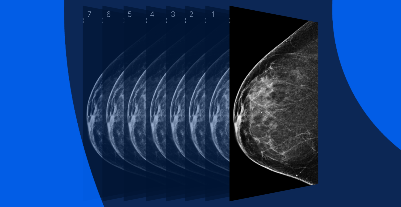
A 3D mammogram is also called digital breast tomosynthesis. The X-ray scanner moves in an arc over the breast, taking multiple images from several different angles. Those X-ray images, called ‘slices’, are digitally combined to provide a comprehensive view of the inside of the breast.
2D mammogram equipment is much more widely available and is often covered by insurance. Not every imaging clinic has 3D mammogram capability, and it can come with additional upfront costs or fees.
However, 3D mammograms present more detailed images of dense breast tissue and can pick up even tinier signs of cancerous growth. Women with dense breasts and a family history of breast cancer may find them helpful. 3D mammograms also produce fewer false positives, meaning fewer patients must be recalled for a second clarifying mammogram.
PocketHealth’s breast health tools provide patients with personalized breast cancer risk assessments and simplified mammogram management. Designed to increase early detection of breast cancer, these tools enable patients to take control of their breast health. Learn more about PocketHealth’s breast health tools here.
Mammograms are used for regular screening purposes and to explore any noticeable external breast abnormalities, such as indentations or lumps, nipple discharge, pain, sores, or thickened skin. Mammograms also measure changes in breast tissue over time, which helps doctors monitor and detect:
Mammograms are also used if a breast biopsy is required, to help position the needle.
The scanning procedure itself is very similar for both 2D and 3D mammograms. You’ll be asked to remove the top half of your clothing and stand in front of the X-ray machine platform, which will be adjusted to your height.
The technician will ask you to place your breast on the X-ray platform and direct you to position your arms, body, and head. A clear plastic plate will compress your breast to the platform while the X-ray scanner takes images. At this point, the technician might ask you to hold your breath or adjust your position slightly to better capture images.
For a 2D mammogram, you’ll be fully repositioned for another scan at a different angle; during a 3D scan, the X-ray scanner will arc over your breast taking multiple image slices. Then the process will repeat for your other breast.
Mammograms are non-invasive imaging exams, but you can do a few things to make your appointment more friction-free.
Here are some suggestions to make your mammogram appointment go more smoothly.
Getting your mammogram is the first step. Next comes obtaining and understanding your results.
Your imaging results are interpreted by a radiologist—an expert in medical imaging—who carefully reviews your scans and provides a detailed report of any findings. This report is then sent to your referring physician, who, with a deeper understanding of your medical history and past exams, can offer further insights and recommendations on next steps in a follow-up appointment.
Turnaround times for imaging results can vary widely depending on the facility and your doctor’s availability. Often, patients wait a week or more and receive their results during a follow-up appointment. If you’d rather not wait anxiously for your results, PocketHealth provides secure access to your mammogram images and report as soon as they’re approved for release by the hospital or imaging clinic. This allows you the opportunity to review your results and prepare questions ahead of your follow-up visit.
Your doctor will discuss your mammogram results with you at a follow-up appointment. To give you an even clearer picture of your mammogram results, PocketHealth provides easy-to-understand definitions and illustrations for complex medical terms—plus an in-depth explanation of your full imaging report. This is paired with highlights of key anatomy in your imaging to help you better comprehend what you’re looking at. This section further explores the categories used to describe mammogram findings and their meanings.
Medical professionals use the Breast Imaging Reporting and Data System (BI-RADS) to sort and describe the findings in mammograms. BI-RADS has 7 categories:
Getting a mammogram can be stressful and uncomfortable, but knowing what to expect can help. Here are a few of the most pressing questions people have about mammograms.
North American health authorities recommend women aged 40 and over who have an average risk of breast cancer, should have screening mammograms every two years. Those with a higher cancer risk might be referred for mammograms more often.
Your insurance might cover most or some of the cost of a 3D mammogram. The exact answer depends on your insurance plan and your location. Ask your insurance provider for details.
No. In both 3D and 2D mammograms, your breast will be pressed between two plates. The pressure will cause some discomfort. If you’re worried about managing pain, ask your doctor about potential pain-reducing options.
It’s not uncommon to be called back for a follow-up mammogram. Being recalled does not mean you have cancer, only that more exploration is required. In fact, less than 10% of patients recalled for another mammogram have signs of cancer. Some of the reasons for mammogram recall include:
A recent radiology study showed that 3D mammograms had a lower recall rate (8.9%) than 2D mammograms (10.3%).
While mammograms are the standard imaging test to monitor breast health, depending on your results you might be sent for other types of imaging, such as a breast MRI, breast ultrasound or molecular breast imaging. This could mean you end up with several different types of breast images to keep track of.
Aileen found it helpful and reassuring to have all her medical images in one secure location, accessible on any device. Having been diagnosed with metastatic breast cancer during the COVID-19 lockdowns, PocketHealth allowed her to share her images and progress when her husband couldn’t accompany her, and gave her a more comprehensive view of her health and progress.
PocketHealth also provides personalized health insights based on the findings in your report to help you stay on top of any next steps. This includes clearly surfacing any follow-up actions found in your report and generating customized questions to ask your doctor so you can make the most of your follow-up appointment.
If you’re located in Ontario, you’ll also have the option to get your breast cancer risk score and determine your eligibility for Ontario Breast Screening Program (OBSP), as part of PocketHealth’s suite of breast health tools. If you’re eligible for OBSP, you can even book your next screening mammogram directly through PocketHealth.
While getting a mammogram may feel overwhelming, better understanding your results and working closely with your healthcare team can give you the best chance of managing any next steps and protecting your health.
Published: December 5, 2024
Trusted by more than 900+ hospitals and clinics.