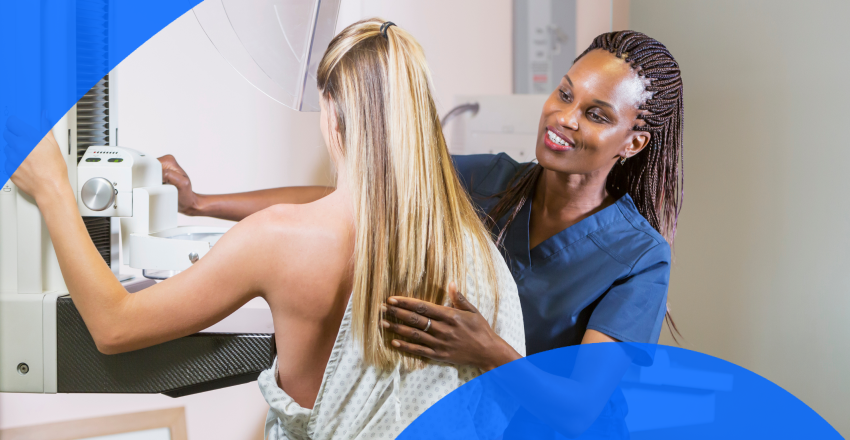
If your doctor has recommended a breast tomosynthesis scan (also called a 3D mammogram), you may have questions about the procedure. This guide will explain this imaging technique, what to expect, and how to prepare for your appointment.
For those looking to take proactive control of their breast health, PocketHealth can help. With secure, easy access to your breast imaging records, PocketHealth allows you to better understand your breast health. In addition, PocketHealth offers personalized estimated breast cancer risk scores to help you and your doctor make informed decisions about preventive measures and future screenings.
Breast tomosynthesis is also known as DBT (digital breast tomosynthesis) or simply a 3D mammogram. This imaging technique is more advanced than typical 2D mammography, which means it may not be available at every imaging clinic. Both mammogram types can detect breast cancer and other breast conditions.
The scan uses low-dose X-rays (electromagnetic radiation) to capture images of internal structures, similar to other types of X-ray imaging. DBT scans are unique because they take several images (called slices) in an arcing pattern, providing far more detail than other X-ray methods.
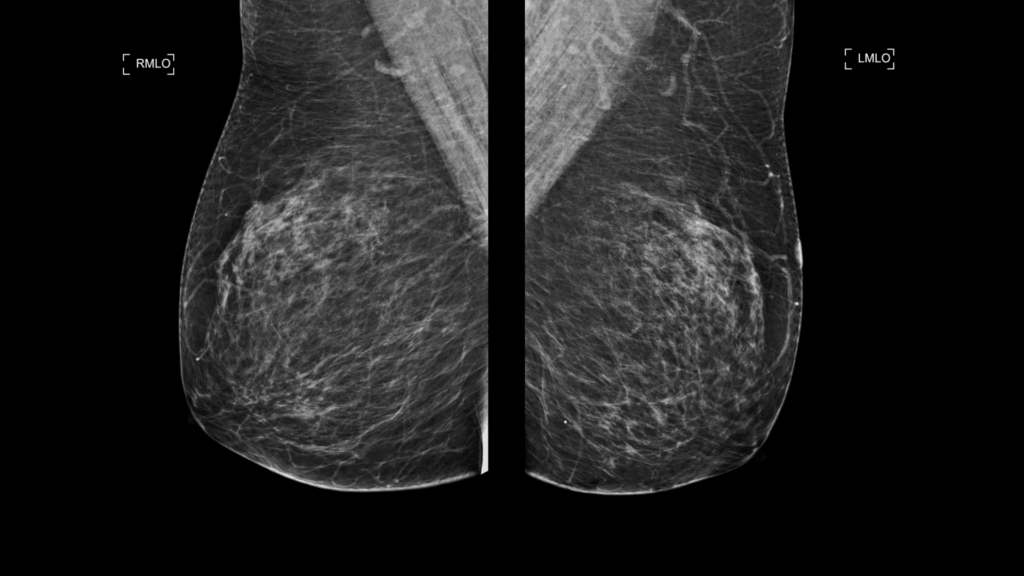
2D mammogram images
Typical mammogram screenings are 2D, with two images taken from different angles for each breast, resulting in only two scans per breast. However, 3D mammograms consist of several images (200 to 300) combined to provide a more detailed view. Because of this, DBT scans result in fewer callbacks, meaning fewer false positives where a finding discovered during the scan is later determined to be benign.
It is not uncommon for 2D mammograms to misidentify normal breast tissue as suspicious for cancer or other conditions. When this happens, patients may require additional scans or biopsies to gather more information. This often proves unnecessary and causes undue stress for the patient. Breast tomosynthesis has lower rates of false positives.
Like 2D mammograms, breast tomosynthesis is a method to help detect breast cancer. It’s especially useful in finding early cases, before patients are more symptomatic. It can be used as a general screening method, which is recommended for all patients assigned female at birth (AFAB) between the ages of 40 and 74. It can also be used for diagnosing symptoms, such as breast pain, lumps etc.
For now, the standard for patients at average risk for breast cancer is still a 2D mammogram. This may change over time, but for most cases, a 2D scan is considered sufficient. A 3D mammogram is recommended in the following conditions:
Breast tomosynthesis scans are performed similarly to a standard 2D scan. The main difference is that the X-ray arm moves in an arc around the breast while capturing images. Here’s what to expect:
Preparation for a DBT scan is the same as for a traditional 2D mammogram. Here are some tips to help you prepare for your appointment.
The mammogram technician is not legally allowed to discuss results during the scan. They can answer questions about the appointment itself, but not about the imaging results. Here are some common questions regarding getting breast imaging results back.
Turnaround times for mammogram results can vary widely depending on the facility and your doctor’s availability. Often, patients wait a week or more and receive their results during a follow-up appointment. With PocketHealth, your imaging results are securely accessible as soon as they’re approved for release by the hospital or imaging clinic. This allows you the opportunity to review your results and prepare questions ahead of your follow-up visit.
A radiologist—an expert in interpreting medical images—will carefully review your breast tomosynthesis and provide a detailed report of any findings. This report is then sent to your referring doctor, who, with a deeper understanding of your medical history, can offer additional insights and recommendations based on the results.
It’s common to find medical terminology confusing. To help you understand your breast imaging results, PocketHealth provides clear definitions and illustrations for complex medical terms—plus an in-depth explanation of your full imaging report. This is paired with highlights of key anatomy in your imaging to help you better comprehend what you’re looking at. Meanwhile, this guide will briefly cover common terminology found in DBT scans.
Common breast conditions that mammograms may reveal include:
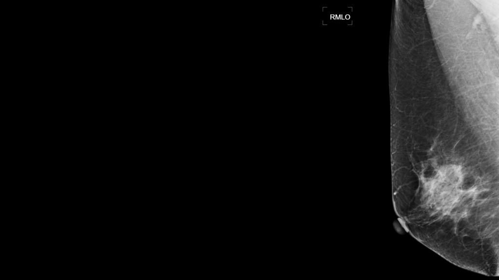
Mammogram showing a benign finding (BI-RADS level 2)
Patients who have undergone 2D mammograms may already be familiar with the Breast Imaging Reporting and Data System (BI-RADS). 3D mammograms use the same system to describe breast imaging results on a scale from 0 to 6, with each level indicating different possible findings or conditions:
Here is a brief overview of frequently asked questions regarding breast tomosynthesis.
Breast tomosynthesis is generally considered an improvement over 2D mammograms. However, because it is relatively new, many facilities do not yet offer it to patients. It can also be more expensive, sometimes involves slightly higher radiation exposure, and may not always be covered by insurance.
3D mammograms can produce higher or lower radiation levels than 2D versions, depending on the equipment being used. As advancements in DBT technology progress, these levels are expected to decrease over time. Regardless, the current radiation exposure is low and considered to be low-risk.
A breast ultrasound does not use X-rays; instead, it uses high-frequency sound waves to create echoes that bounce off internal structures and capture images. Ultrasounds and mammograms work well together, providing additional imaging when needed. 3D mammograms are effective at detecting early signs of breast cancer or other symptoms. However, if more clarity is required on DBT findings, breast ultrasounds can provide better details, such as determining whether a lump is a fluid-filled cyst or a solid mass.
Like breast ultrasounds, breast MRIs (magnetic resonance imaging) are a useful complement to a 3D mammogram. This technique uses magnets and radio waves to produce 3D images of the patient’s internal anatomy. Some patients may require an MRI instead of a mammogram, particularly those at higher risk for breast cancer who need more sensitive imaging. However, more commonly, MRIs are used as a supplement to mammograms. This can include situations where more detail is needed on an abnormal finding, when the patient has breast implants that may obstruct views and when the patient has very dense breasts. Breast MRIs are highly detailed, making them a strong screening and diagnostic tool, but they can also lead to false positives, as they may detect benign findings more frequently than other imaging methods.
Given the routine nature of breast imaging, tracking changes over time is essential. PocketHealth provides patients with personalized breast cancer risk assessments and simplified mammogram management. Designed to increase early detection of breast cancer, these tools enable patients to take control of their breast health. Learn more about PocketHealth’s breast health tools here.
Having secure access to all your imaging records in one place allows you to be actively involved in your health journey. PocketHealth makes it simple to keep track of your medical imaging results, including your breast scans. All of your images and reports are permanently available in one secure location and can be accessed online—anytime, anywhere. Reports can also be easily shared with other members of your care team, and you can even print and store these reports for your own use. When used in conjunction with your medical provider’s professional advice, it is a powerful tool to better understand your imaging results.
PocketHealth also provides personalized health insights based on the findings in your report to help you stay on top of any next steps. This includes clearly surfacing any follow-up actions found in your report and generating customized questions to ask your doctor so you can make the most of your follow-up appointment. For Mary, accessing her breast imaging through PocketHealth helped her catch the medical recommendation for more frequent mammogram screenings, something her clinic had missed.
While breast imaging may feel overwhelming, better understanding your results and working closely with your healthcare team can give you the best chance of managing your condition and protecting your health.
Published: January 5, 2025
Trusted by more than 900+ hospitals and clinics.