Mammograms have been an important screening and diagnostic method for years. Most people are familiar with them, but 3D mammograms are a little different. This guide will discuss what makes this imaging technique unique, who benefits most from it and what to expect during the scan.
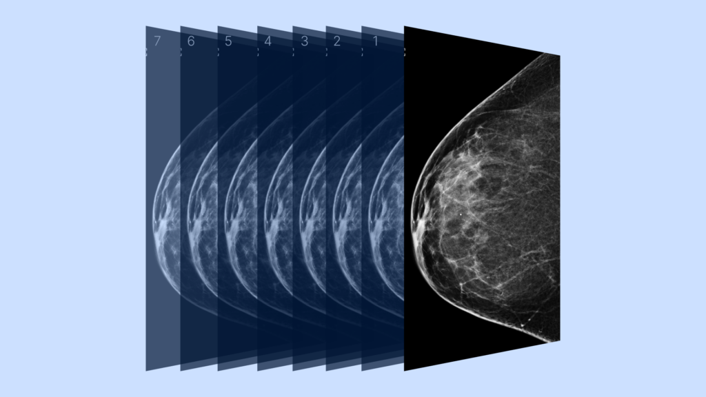
A simplified example of how a 3D mammogram image is built using many image slices taken at different angles, sourced from the National Cancer Institute via National Institutes of Health Image Gallery.
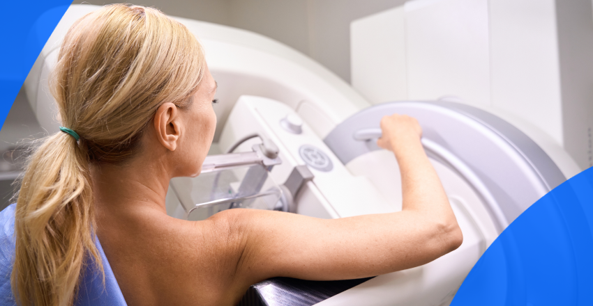
A mammogram is an X-ray that uses a very low radiation dose to examine breast tissue. These images can reveal any abnormal changes before they can be felt in a traditional breast exam.
In a 3D mammogram, the X-ray moves in an arc over the breast, taking multiple pictures from several different angles. Those images, called slices, are combined, providing a more complete view of the breast and making it easier to spot any irregularities. 3D mammograms are highly effective. A 2023 study of more than 1 million women showed that they led both to a higher rate of cancer detection and a lower rate of recalls for further imaging.
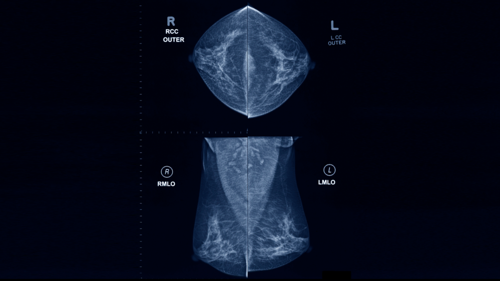
A 2D mammogram image
A traditional 2D mammogram provides 2 images of each breast (from the top and the side). 3D mammograms provide hundreds of thinly sliced images, which come together to create a highly detailed 3D view of the whole breast.
Mammograms are also the only way to assess breast density, which is important because dense breasts have increased cancer risks and can make abnormalities harder to spot. 3D mammograms are more effective for dense breasts because physicians can see the dense areas from several different angles.
A traditional 2D mammogram presents two overlapping images. That overlap can obscure potential abnormalities, causing them to be missed at the earliest stages. It’s also possible for overlapping tissue to make benign spots look abnormal, which then requires you to have further imaging done for clarification.
Some patients may benefit more from a 3D mammogram instead of a 2D one, especially if they have a higher risk of or history of breast cancer. Some advantages include:
According to this 2023 study, 3D mammograms detect cancer in 5.3 out of 1000 cases, whereas traditional mammograms found cancer in 4.5 out of 1000. That’s a significant difference when it comes to early detection and treatment. Studies have also shown that 3D mammograms detect more kinds of cancer than regular 2D mammograms.
Medical bodies suggest different starting ages, but in 2023, the USPSTF issued a draft recommendation that women start regular screening mammograms every other year beginning at age 40. At 55, you can scan every year in the U.S.
Should you have a family health history of breast cancer, you should begin your screening sooner, and consider both mammograms and MRIs. Always discuss your risk factors with your physician so you can explore the best scan options for your particular needs.
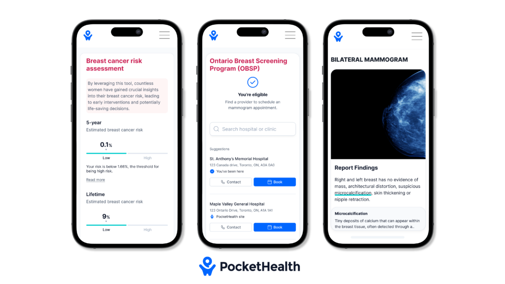
PocketHealth recently launched breast health tools that provide patients with personalized breast cancer risk assessments and simplified mammogram management. Designed to increase early detection of breast cancer, these tools enable patients to take control of their breast health. Learn more about PocketHealth’s breast health tools here.
While a 3D mammogram only requires a short appointment, there are some steps you can take to prepare. Here is a quick overview.
Ideally, you’ll want to make these preparations before your actual appointment.
Here are some day-of tips for your appointment:
As with all medical procedures, there are pros and cons when getting a 3D mammogram.
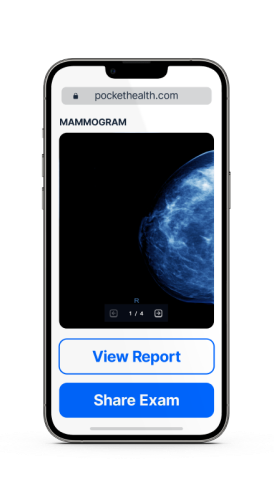
Mammogram result turnaround times depend on the schedules of the radiologist and your doctor. They can take up to a week or more, especially if your doctor has scheduled a follow-up appointment to discuss your results. For patients wanting faster access, you can view your images and report through PocketHealth, often before your follow-up appointment.
PocketHealth allows you to access, view and store your medical images and health information all in one secure location. You can also use PocketHealth to share images, which is helpful if other medical providers need to review your mammograms.
Your imaging results are interpreted by a radiologist—an expert in medical imaging—who carefully reviews your scans and provides a detailed report of any findings. This report is then sent to your referring physician, who, with a deeper understanding of your medical history and past exams, can offer further insights and recommendations on next steps in a follow-up appointment.
Turnaround times for imaging results can vary widely depending on the facility and your doctor’s availability. Often, patients wait a week or more and receive their results during a follow-up appointment. With PocketHealth, your imaging results are securely accessible as soon as they’re approved for release by the hospital or imaging clinic. This allows you the opportunity to review your results and prepare questions ahead of your follow-up visit.
To help you understand your mammogram results, PocketHealth provides clear definitions and illustrations for complex medical terms—plus an in-depth explanation of your full imaging report. This is paired with highlights of key anatomy in your imaging to help you better comprehend what you’re looking at.
Below are explanations of some of the conditions that mammograms typically scan for. While it can be easy to worry if you see these results in your report, just remember that it is very common for abnormalities to be benign. Your doctor will guide you on any additional testing or steps. Here are some helpful terms to know:
In a normal breast image, the background will be black while the breast tissue shows up in shades of gray. Dense tissue and glands will be white, whereas abnormalities like the ones listed above will have unique appearances. For example:
Some of these abnormalities will suggest further testing, which might include another mammogram or a biopsy.
Doctors use a standard vocabulary to describe mammogram results, called the Breast Imaging Reporting and Data System (BI-RADS). BI-RADS has 7 numbered categories:
Regular mammograms are vital for breast cancer detection and prevention. Mammograms of all types have life-saving potential since they can detect signs of breast cancer before you can feel them. Discuss 3D mammography with your physician to see if it’s the right choice for you.
It’s important to stay both informed and proactive when it comes to maintaining your breast health. PocketHealth makes it simple to keep track of your medical imaging results. All of your images and reports are permanently available in one secure location and can be accessed online—anytime, anywhere. Reports can also be easily shared with other members of your care team, if needed. When used in conjunction with your medical provider’s professional advice, it is a powerful tool to better understand your imaging results.
PocketHealth also provides personalized health insights based on the findings in your report to help you stay on top of any next steps. This includes clearly surfacing any follow-up actions found in your report and generating customized questions to ask your doctor so you can make the most of your follow-up appointment. For Mary, being able to review and compare her breast imaging led to the discovery that she was recommended to have these scans more frequently—something her primary doctor did not catch.
If you’re located in Ontario, you’ll also have the option to get your breast cancer risk score and determine your eligibility for Ontario Breast Screening Program (OBSP), as part of PocketHealth’s suite of breast health tools. If you’re eligible for OBSP, you can even book your next mammogram directly through PocketHealth.
While getting any mammogram imaging may feel overwhelming, better understanding your results and working closely with your healthcare team can give you the best chance of managing your condition and advocating for your health.
Published: September 5, 2023
Trusted by more than 900+ hospitals and clinics.