
Second only to skin cancer, breast cancer is the most commonly diagnosed cancer among individuals assigned female at birth (AFAB) in the United States. Although it is most frequently identified in those who are middle-aged and older, it can also occur in younger patients. This article presents an overview of the condition, examines staging and diagnostic methodologies and outlines current treatment approaches.
Breast cancer occurs when cells in the breast divide and grow uncontrollably, often forming tumors that can impact the body’s function and overall health. While the majority of breast cancer patients are AFAB, those assigned male at birth (AMAB) can also be affected, though this is rare.
Breast cancer can develop in specific parts of the breast, and its origin often determines how it is classified. Some examples include:
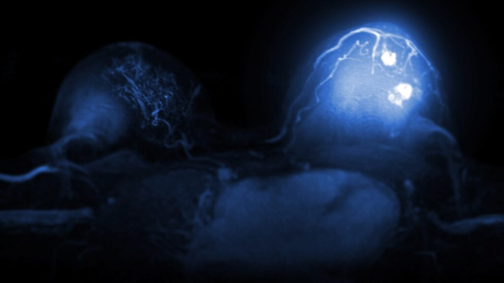
A breast MRI with contrast
There are multiple methods to diagnose breast cancer. Here is a brief overview of each.
Some patients discover breast lumps or irregularities during a self-breast exam, while others may receive a clinical breast exam as part of routine medical care or screenings. The breasts, nipples, underarms and collarbone areas are assessed for abnormalities, lumps or other changes that could indicate a tumor. It’s worth noting that most breast lumps are benign. If a lump is found, additional testing is typically performed.
There are multiple imaging methods that can help diagnose breast cancer, including:
One of the most definitive ways to diagnose breast cancer is with a biopsy. This involves collecting a small sample of breast tissue to be analyzed in a laboratory for possible cancer cells. It’s typically done using a thin, hollow needle, guided by an MRI or other breast imaging. A fine-needle biopsy may be used for smaller samples, while a core needle biopsy is preferred for collecting larger ones. In rare cases, a surgical biopsy may be performed if the doctor wants to remove the entire lump for analysis. Nearby lymph nodes may also be biopsied to determine whether the cancer has spread.
Often, during the biopsy, a small metal marker is placed in the area where the tissue sample was taken. Because this marker shows up on breast imaging, it serves as a helpful way to monitor and evaluate the specific area for any future changes.
Breast cancer staging is an important part of both diagnosis and treatment. The tissue sample taken from the biopsy is evaluated not only for cancer cells, but also for details on subtypes and if there are hormone receptors or proteins that are feeding the cancer. This is often called a biomarker test. There are other methods to help stage breast cancer, including:
Not all patients will require all of these tests to determine staging. Often the physician will select the ones most applicable to reach a staging diagnosis. Here are the stages to be aware of:
How breast cancer is treated depends on factors such as stage, type or subtype and grade. Cancer grade is determined by studying biopsied cancer cells and may indicate how fast the cancer is growing. Grade one means the cancer cells look similar to normal cells, suggesting slower growth. Grades two and three are more abnormal in appearance, suggesting faster growth. All of these aspects—including biomarkers and hormone receptors—are taken into account when deciding on treatment. Here are some common treatment methods.
The type of breast cancer surgery will depend on factors such as grading, staging and cancer type. Common surgical methods include:
This treatment uses powerful pharmaceutical drugs that may be administered intravenously or in pill form. Chemotherapy is usually given after the surgical removal of the tumor, though it may be used beforehand if the doctor wants to shrink the tumor first. It can also help lower the risk of cancer recurrence or the spread of cancer cells to the lymph nodes. Common side effects may include nausea, fatigue, temporary hair loss and an increased risk of infection.
Because some types of breast cancer feed on hormones like estrogen and progesterone, hormone therapy works by blocking these hormones, preventing cancer cells from being fueled by them. Like chemotherapy, hormone therapy is usually given after surgery. It helps reduce the risk of cancer recurrence and is often taken for up to 5 years or more. Possible side effects may include vaginal dryness, hot flashes, night sweats, blood clots and bone thinning.
Targeted therapy uses medications that attack specific substances cancer cells rely on to grow, such as the protein HER2. By targeting and blocking HER2, these drugs can cut off the cancer cell’s fuel source, preventing further growth. The type of medication used depends on which substances are being targeted. This therapy may be given before breast cancer surgery to help shrink the tumor, or afterward to reduce the risk of recurrence.
Immunotherapy uses special medications to boost the body’s own immune system, helping it more effectively recognize and destroy cancer cells. Since cancer can often hide from the immune system, this treatment helps the body detect and defend against those cells more successfully.
Radiation therapy uses strong energy beams, such as protons or X-rays, to destroy cancer cells—often after surgery. There are two types of radiation therapy: external (called external beam radiation therapy) and internal (called brachytherapy). Radiation is often associated with lower rates of cancer recurrence, as it targets and destroys remaining cancer cells after surgery. Common side effects include fatigue, inflammation and sunburn-like skin symptoms.
Your breast imaging results are interpreted by a radiologist—an expert in medical imaging—who carefully reviews your scans and provides a detailed report of any findings. This report is then sent to your referring physician, who, with a deeper understanding of your medical history and past exams, can offer further insights and recommendations on next steps in a follow-up appointment.
Turnaround times for breast imaging can vary widely depending on the facility and your doctor’s availability. Often, patients wait a week or more and receive their results during a follow-up appointment. With PocketHealth, your imaging results are securely accessible as soon as they’re approved for release by the hospital or imaging clinic. This allows you the opportunity to review your results and prepare questions ahead of your follow-up visit.
To help you understand your breast imaging results, PocketHealth provides clear definitions and illustrations for complex medical terms—plus an in-depth explanation of your full imaging report. This is paired with highlights of key anatomy in your imaging to help you better comprehend what you’re looking at.
Here are some common questions regarding breast cancer.
Understanding breast cancer risk can be important, as high-risk patients may have increased screening recommendations. Several factors may influence a patient’s risk of developing breast cancer, including:
Mutations in the BRCA1 and BRCA2 genes can increase the risk of breast cancer. For patients who have already undergone genetic testing and haven’t developed breast cancer, it may be worth discussing with their doctor whether they should have more frequent cancer screenings to detect potential cancer cells early.
For patients already diagnosed with breast cancer, genetic testing can help guide treatment decisions and offer insight into whether there is a genetic aspect to their diagnosis. This information can also be useful for family members, allowing them to discuss with their medical provider whether they should be tested for mutations.
Breast cancer recurrence happens when a previous breast cancer returns. It isn’t a new case of cancer—rather, it means that cancer cells from the original diagnosis were not completely eliminated. Over time, these cells can divide and spread again, leading to a recurrence.
All cancer patients face some risk of recurrence, though in most cases, it does not occur. Recurrence rates depend on the original cancer stage, how it was treated and other individualized factors.
Breasts are composed of both fatty tissue and fibroglandular tissue. When there is less fatty tissue compared to fibroglandular tissue, they may be categorized as dense. Dense breasts can make it more difficult to detect lumps and irregularities during imaging tests, especially a mammogram. They can also increase a patient’s risk of developing breast cancer.
Over 50% of AFAB patients over 40 have dense breasts, making it a common occurrence. A mammogram or other breast screening should indicate whether or not breasts are dense so patients can be aware of any increased risks. If there are concerns or questions, asking a medical provider about next steps may be a good course of action. They may recommend more detailed breast imaging, such as an MRI.
Regardless of the stage, a breast cancer diagnosis can feel overwhelming. It’s important to remember that you’re not alone and that there are support systems and resources available. Your medical provider will likely be able to connect you with local options. There are also many online support groups and informational resources. Here are some breast cancer organizations that some may find helpful:
PocketHealth makes it simple to keep track of your breast scans. All of your images and reports are permanently available in one secure location and can be accessed online—anytime, anywhere. Reports can also be easily shared with other members of your care team, if needed. When used in conjunction with your medical provider’s professional advice, it is a powerful tool to better understand your health. For Aileen, easy access to her scans also allowed her to keep her husband informed throughout her metastatic breast cancer treatment.
PocketHealth also provides personalized health insights based on the findings in your report to help you stay on top of any next steps. This includes clearly surfacing any follow-up actions found in your report and generating customized questions to ask your doctor so you can make the most of your follow-up appointment.
While a breast cancer diagnosis is stressful and overwhelming, better understanding your results and working closely with your healthcare team can give you the best chance of managing your condition and protecting your health long term.
Published: April 24, 2025
Trusted by more than 900+ hospitals and clinics.